
Image
|
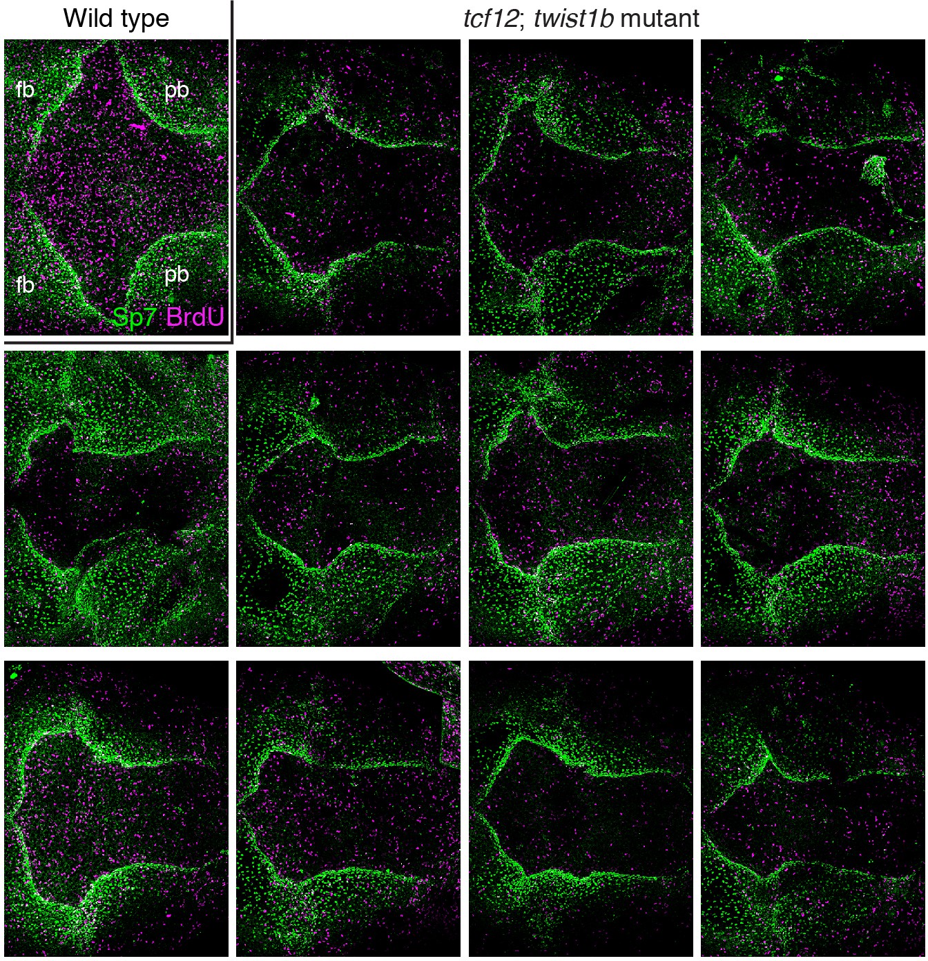
Figure Caption
Fig. 5-S2
Additional examples of BrdU and Sp7 staining in tcf12; twist1b mutant zebrafish.
Dorsal views of 9 mm zebrafish skullcaps stained with Sp7 antibody (green) and BrdU (magenta) show an array of altered bone shapes in mutants compared to wild type. fb, frontal bone; pb, parietal bone.
Figure Data
Acknowledgments
This image is the copyrighted work of the attributed author or publisher, and
ZFIN has permission only to display this image to its users.
Additional permissions should be obtained from the applicable author or publisher of the image.
Full text @ Elife