
|
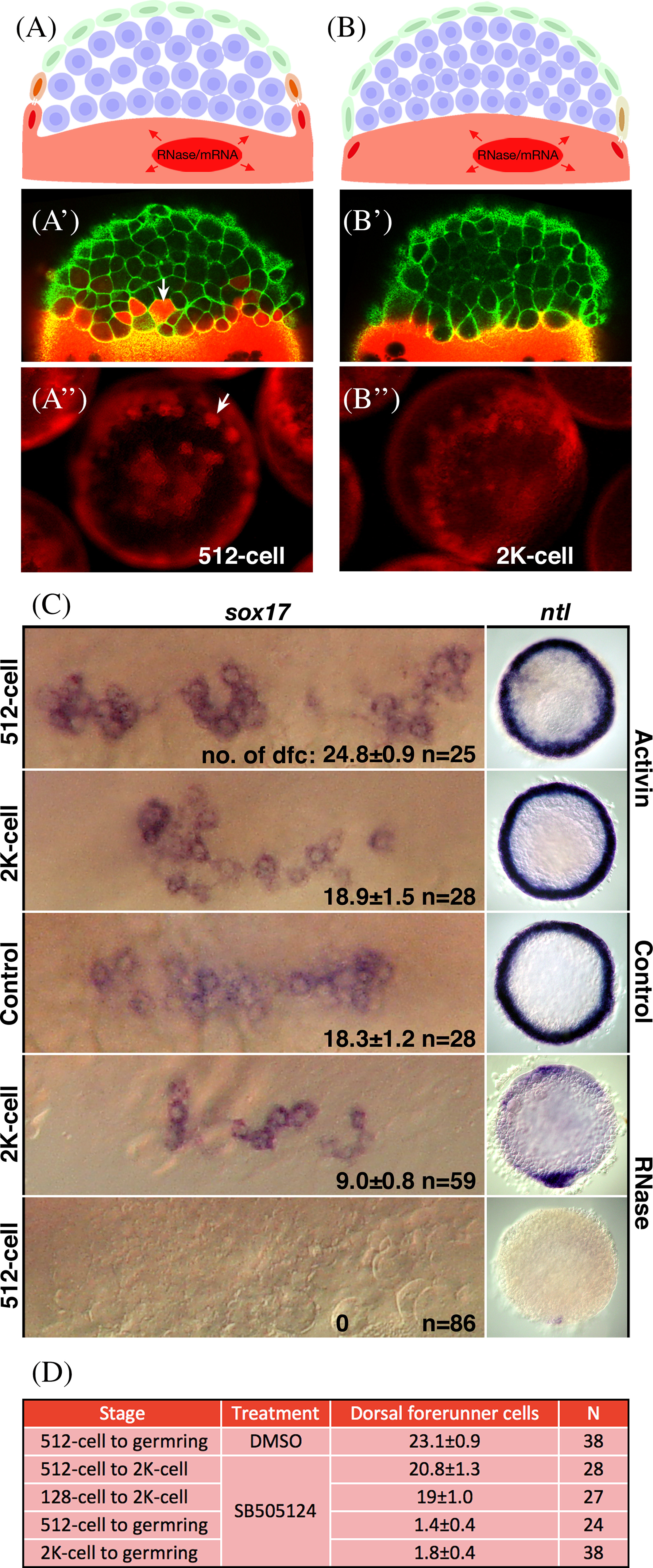
Fig. 6
Yolk cell cytoplasm is necessary for forerunner fate. A,B: Experimental design and examples. Injection of lineage tracer dye into the yolk cell at the 512‐cell stage (3 h, A), when Wilson cells are present, the dye not only incorporates into the yolk cell, but also into Wilson cells and the cells that they are bridged to (white bars). Injection into the yolk cell at the 2K‐cell stage (3.5 h, B), after Wilson cells collapse to form the yolk syncytial layer, shows dye only incorporates into the yolk cell or very weakly into enveloping layer cells that were once coupled to a Wilson cell. A′,B′: Confocal images of transgenic embryos carrying Tg(β‐actin: utrophin‐GFP) to outline cells in green and injected with red lineage tracer as for A and B. White arrow indicates one marked cell in the 512‐cell stage embryo, there are many others in this plane of focus, but no marked cells in the 2K‐cell stage embryo. A″,B″: Images of experimental embryos injected as for A and B, and, in this example, with RNaseA. White arrow indicates one marked cell in the 512‐cell stage embryo, there are many others in this embryo. Robust labeling around the blastoderm margin in the 2K‐cell stage embryo is in the yolk syncytial layer. C: Results of injections with Activin mRNA and RNaseA. Views for sox17 are face views at higher magnification and views for ntl are animal pole views, dorsal oriented down. Injection of Activin mRNA at the 2K‐cell stage had no effect on the number of sox17 positive dorsal forerunner cells or expression of ntl in the presumptive mesendoderm at 50% epiboly, but injection at the 512‐cell stage (3 h) caused an approximate 40% increase in the number of dorsal forerunner cells and a marked expansion of presumptive mesendoderm. Injection of RNaseA at the 2K‐cell stage (3.5 h) reduced the number of sox17 positive dorsal forerunner cells by approximately 50% and decreased the expression of ntl in the presumptive mesoderm at 50% epiboly (5.3 h) all but dorsally, but injection at the 512‐cell stage (3 h) abolished the dorsal forerunner cells altogether and almost all presumptive mesendoderm except a smidgen dorsally. D: Effects of inhibiting the Nodal‐signaling pathway with SB‐505124 or DMSO (control) at the times indicated. Dorsal forerunner cells were visualized and counted at 50% epiboly using sox17 expression as shown above. Their numbers are shown as average ± SE; N is number of embryos. There is little effect on blocking the Nodel signaling pathway between the 512‐cell to 2K‐cell stage, but blocking after the 2K‐cell stage prevents dorsal forerunner cells from forming.