
|
Fig. 5
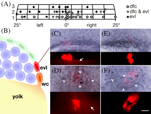
Forerunner cells originate from enveloping layer cells that share lineage with Wilson cells. A: Gastrula fate map for the dorsal forerunner cells. The x‐axis is distance in degrees from dorsal; y‐axis is in cell diameters from the margin. Each grid is roughly the area of a single enveloping layer cell and each symbol plots the location of a single enveloping layer subclone, typically the average location of two cells. B–F: Forerunner cells derive from enveloping layer cells coupled to a Wilson cell. B: Experimental design. Injection into an enveloping layer cell (evl) at the 512‐cell stage (3 h) to determine whether it is dye‐coupled through a midbody (white bars) to a Wilson cell (wc) and indirectly to the yolk cell. Enveloping layer cells are green, deep cells are blue, Wilson cells have orange nuclei and the yolk is yellow. C: An enveloping layer cell (after its division) that was coupled to a 512‐cell stage (3 h) Wilson cell. Arrow indicates dye coupling to yolk cell. D: At 60% epiboly (6.5 h), this clone contributed progeny to the forerunner cluster (arrowheads). Of 42 such clones, 15 produced unrestricted clones that included forerunners, and 22 produced restricted forerunner clones; the remaining 5 produced restricted periderm clones. Arrow indicates dye coupling to yolk cell. E: An enveloping layer cell (after its division) not coupled to a 512‐cell stage (3 h) Wilson cell. F: At 60% epiboly (6.5 h), this clone contributed progeny to enveloping layer cells over the forerunner cluster. Of 38 such clones, 36 produced restricted periderm clones; the remaining 2 produced unrestricted clones that included forerunner derivatives.