
|
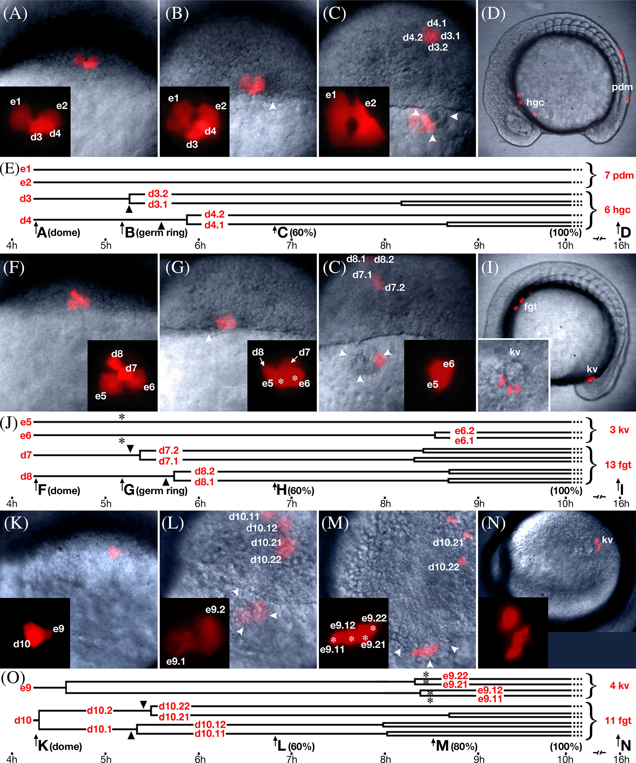
Fig. 2
Lineage analysis of dorsal marginal enveloping layer cells. Selected frames from multi‐plane video time‐lapse recordings of embryos containing dorsal enveloping layer subclones. No entire frame has the whole clone in focus; we selected frames that were approximately the best for showing the multiple cells. Enveloping layer cells labeled are with e numbers; deep domain cells, with d numbers. For this analysis, 40 clones were analyzed in 10 planes of focus. No example is shown for unrestricted enveloping layer subclones where both periderm and forerunner fates were produced (six cases). A–E: Example of an enveloping layer subclone restricted to periderm fate, representative of 17 subclone pairs. A: Beginning of recording at doming (4.3 h), when there were two enveloping layer cells and two deep cells (see Supplementary Movie S1). B: At germring (5.7 h), the enveloping layer cells retained their morphology while the two deep cells began to internalize. Nearby, the nascent forerunner cluster was visible (arrowheads). C: At 60% epiboly (6.5 h), the enveloping layer subclone moved over the forerunner cluster whereas the deep subclone ingressed into the prechordal mesendoderm and moved toward the animal pole. Insets show magnified view of enveloping layer subclone. D: At 14 somites (16 h), the enveloping layer subclone was over the tail and became periderm (pdm); the deep subclone was near the head and became hatching gland (hgc). E: Lineage diagram of subclones in A–D; e‐ and d‐numbers refer to cells in above panels; arrows and block letters in timeline refer to panels above and stages are in parentheses. Arrowheads indicate time of deep cell ingression. F–J: Example of enveloping layer subclone restricted to forerunner fate, entering the forerunner cluster at the onset of gastrulation, representative of 6 subclone pairs (see Supplementary Movie S2). No example is shown for cells entering the forerunner cluster both at the onset and midway through gastrulation (4 cases). F: Beginning of recording at doming (4.3 h), when there were two enveloping layer cells and two deep cells. G: At germring (5.7 h), the enveloping layer cells (asterisks) round up as the deep cells ingressed into the mesendodermal layer. H: At 60% (6.5 h) epiboly, the enveloping layer subclone now with changed morphology enters the forerunner cluster; above, the deep subclone has moved into prechordal mesendoderm. Insets show magnified view of enveloping layer subclone. I: At 14 somites (16 h), the enveloping layer clone was in Kupffer's vesicle (kv), whereas the deep subclone was rostral in the foregut (fgt). Additional inset shows magnified view of three cuboidal cells in Kupffer's vesicle. J: Lineage diagram of subclones in F–I, labeled as in E; asterisks indicate time of change from enveloping layer to forerunner cell morphology. K–O: Example of enveloping layer subclones restricted to forerunner fate, entering the forerunner cluster midway through gastrulation, representative of seven subclone pairs. K: Beginning of recording at doming (4.3 h), there was one enveloping layer cell and one deep cell. L: At 60% epiboly (6.5 h), the enveloping layer subclone was over the forerunner cluster but retains an enveloping layer morphology, and the deep subclone has ingressed into the prechordal mesendoderm and moved animalwards. M: At 80% epiboly (8 h), the enveloping layer cells (asterisks) rounded up and clearly displayed forerunner morphology. N: At 14 somites (16 h), the enveloping layer clone was in Kupffer's vesicle (kv); and the deep subclone was in the foregut (fgt), but not visible in this view. Additional inset shows magnified view of three of the four cuboidal cells in Kupffer's vesicle. O: Lineage diagram of subclones in K–O, labeled as in E and J.