
|
Fig. 1
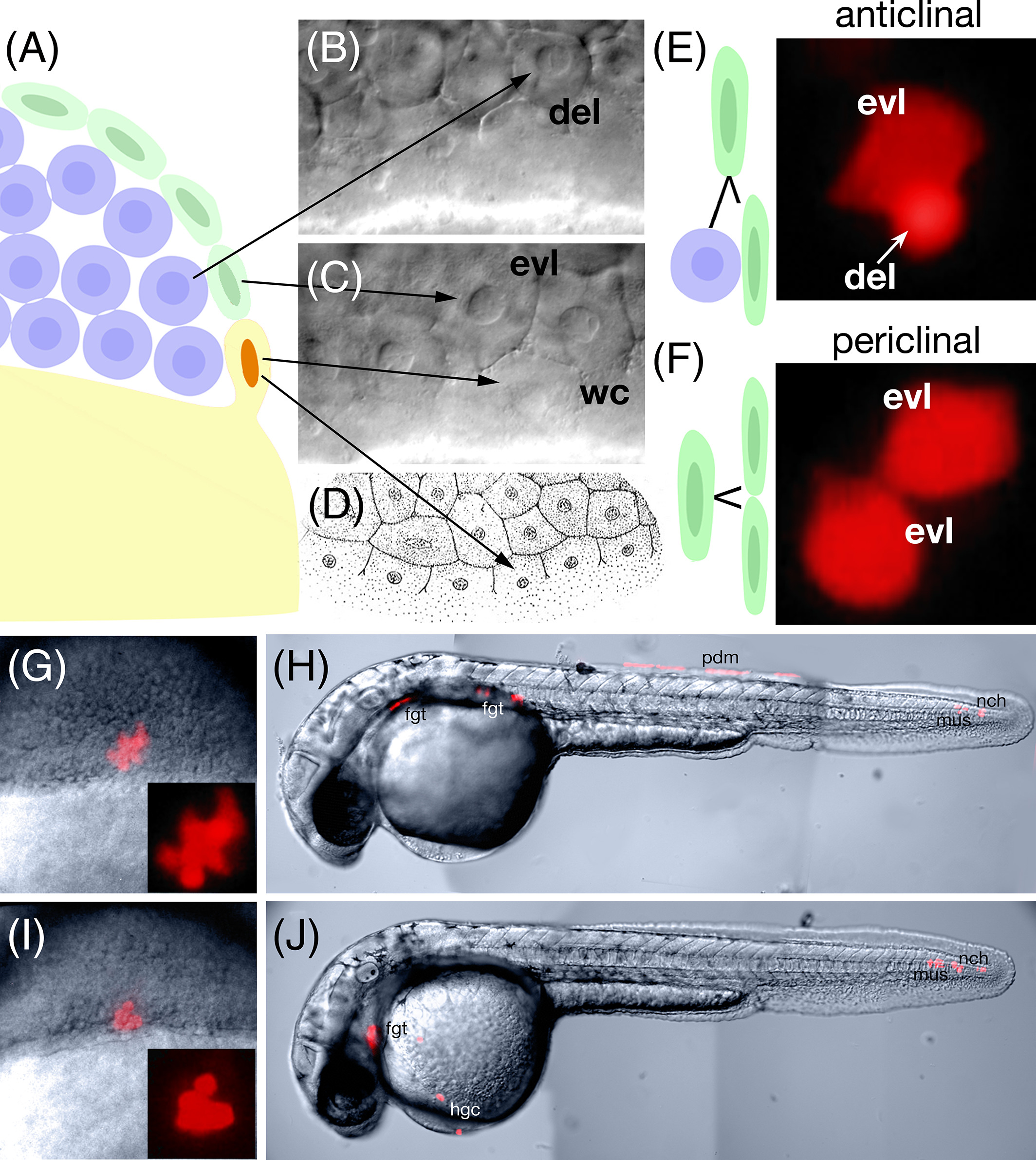
Enveloping layer clonal fates inconsistent with the zebrafish fate map. A: Schematic of the three domains of the blastoderm at the mid blastula transition (3 h): exterior enveloping layer cells (green), deep domain cells (blue), and Wilson cells (orange nuclei), which are cytoplasmically continuous with the yolk cell (yellow). B–D: Deep (B) and superfical (C) focal plane of the blastoderm showing the three cell types. Abbreviations: evl, enveloping layer; wc, Wilson cell; del, deep cell. (D) Figure 22 from plate 90 (Supplementary Figs. S1 and S2) of H. V. Wilson (1889) showing a surface view of the blastoderm edge. Marginal cells cytoplasmically continuous with the yolk, which we term Wilson cells in this study, will after one or two more divisions collapse into the yolk cell to become the yolk syncytial layer. E,F: Lineage relationships of enveloping layer cells before the 4K‐cell stage. Enveloping layer cells can divide (E) anticlinally to produce an evl‐del sibling pair or (F) periclinally to produce and evl‐evl sibling pair. The evl‐del sibling pair will go on to produce a subclone of enveloping layer cells and a subclone of deep cells whereas the evl‐evl sibling pair will go on to produce a clone exclusively of enveloping layer cells. Deep cells can be easily identified by their smaller apparent size, spherical shape and intense fluorescence compared with the larger, flatter and polygonal‐shaped enveloping layer cells whose fluorescence is more diffuse due their thinner dimension in face view. Fluorescent images to the right show examples from our documentation. G–J: Examples of dorsal marginal clones derived from anticlinal divisions of labeled enveloping layer cells. G,H: Example of a clone where (G) at shield stage (6 h) there was a subclone of enveloping layer cells (less intense polygonal cells) and a subclone of deep cells (very intense round cells). Later in the 30‐h embryo (H), there was posterior periderm (pdm) derived from the enveloping layer subclone as well as nearby tail mesoderm notochord (nch) and muscle (mus), not normally derived from dorsal marginal deep cells, but typical derivatives of dorsal forerunner cells. In the head there was also foregut endoderm (fgt) a typical derivative of dorsal marginal deep cells. I,J: Example of a clone where (I) at shield stage, the enveloping layer subclone was no longer apparent and the clone appeared to be composed of only deep cells (intensely labeled round cells). By 30 h (J), where the enveloping layer subclone should have been, there was tail mesoderm: notochord and muscle. In the head there were the normal dorsal mesodermal fates: hatching gland cells (hgc) and foregut endoderm.