
|
Fig. S1
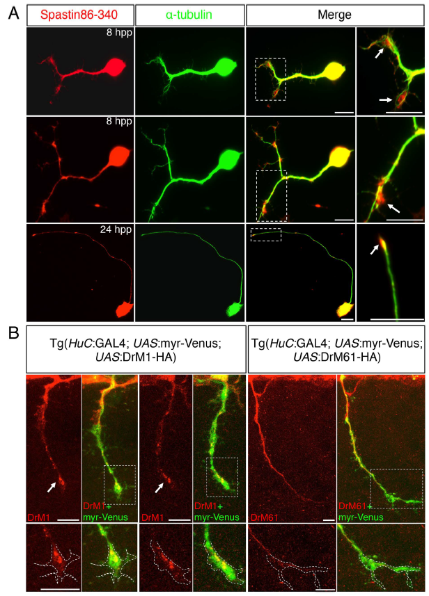
Subcellular distribution of DrM1 and DrM61 spastin isoforms in zebrafish spinal neurons. (A) Primary cultures of zebrafish spinal neurons were immunolabelled 8 or 24 hour post-plating (hpp) with spastin 86-340 (red) and α-tubulin (green) antibodies. Arrows indicate spastin enrichment in growth cones. Right panels are higher magnifications of the boxed region in the corresponding left panel. (B) Tg (HuC:Gal4; UAS:myr-Venus; UAS:DrM1) and Tg (HuC:Gal4; UAS:myr-Venus; UAS:DrM61) embryos were immunolabelled at 28-hpf with HA and GFP antibodies. Arrows point at DrM1 spastin enrichment in SMN growth cones. Bottom panels represent higher magnifications of the boxed region in the corresponding upper panel. Dashed lines in bottom panels delineate the growth cone area. (A-B) Scale bars: 10 μm.