
|
Fig. 5
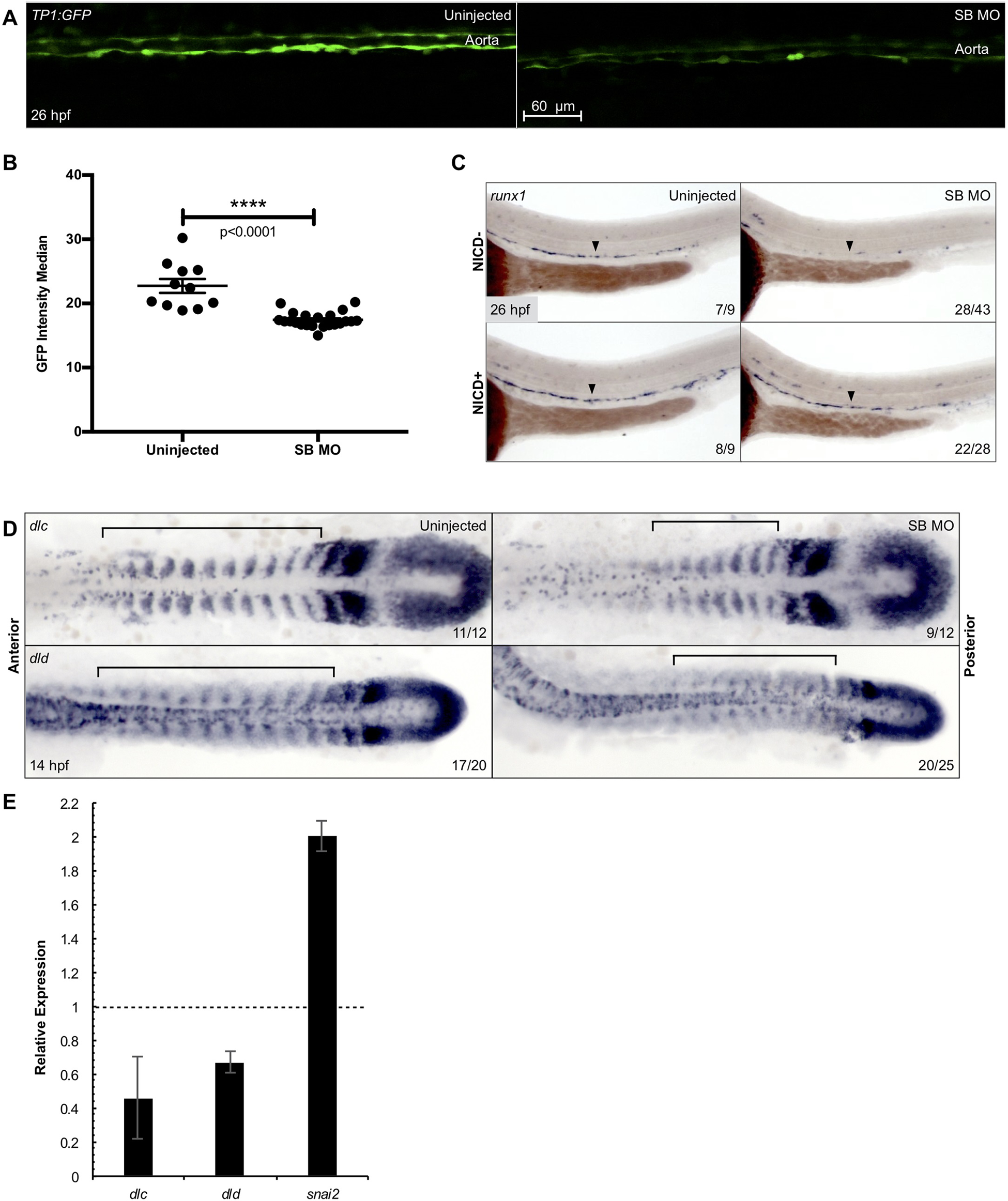
Snai2 SB morphants have defective Notch signaling.
Aortic Notch activity was assessed by confocal microscopy of the Notch reporter Tg(TP1:GFP) (A). Median fluorescence intensity was calculated by the surfaces feature of Imaris and was graphed and statistically analyzed by a non-parametric t-test on Prism (B). Error bars are SEM. Using a combination of Tg(kdrl:miniGal4) and Tg(UAS:NICD-myc), we saw that ectopically activating Notch signaling within the endothelium was sufficient to rescue expression of the HSC marker runx1 in morphant embryos (C). Black arrowheads point to the middle of the aortic runx1 expression. Analysis by WISH displays that expression of the Notch ligands dlc and dld is decreased in morphants, especially within the more anterior somites (D). Black brackets are provided to highlight the differences in staining. This decrease was further confirmed by qPCR in somitic, GFP+ cells sorted from morphant Tg(actc1b:GFP) embryos as compared to uninjected siblings (E). Snai2 relative expression is included for comparison, since the misspliced transcript is consistently elevated in SB MO injected embryos. Error bars are calculated from technical replicates. Numbers in the lower right-hand corner of each image depict the number of embryos with the phenotype pictured out of the total number of embryos assayed in each condition.