
|
Fig. 3
mnr2b-ABNs in r5 and r6 Form Convergent Efferent Projections
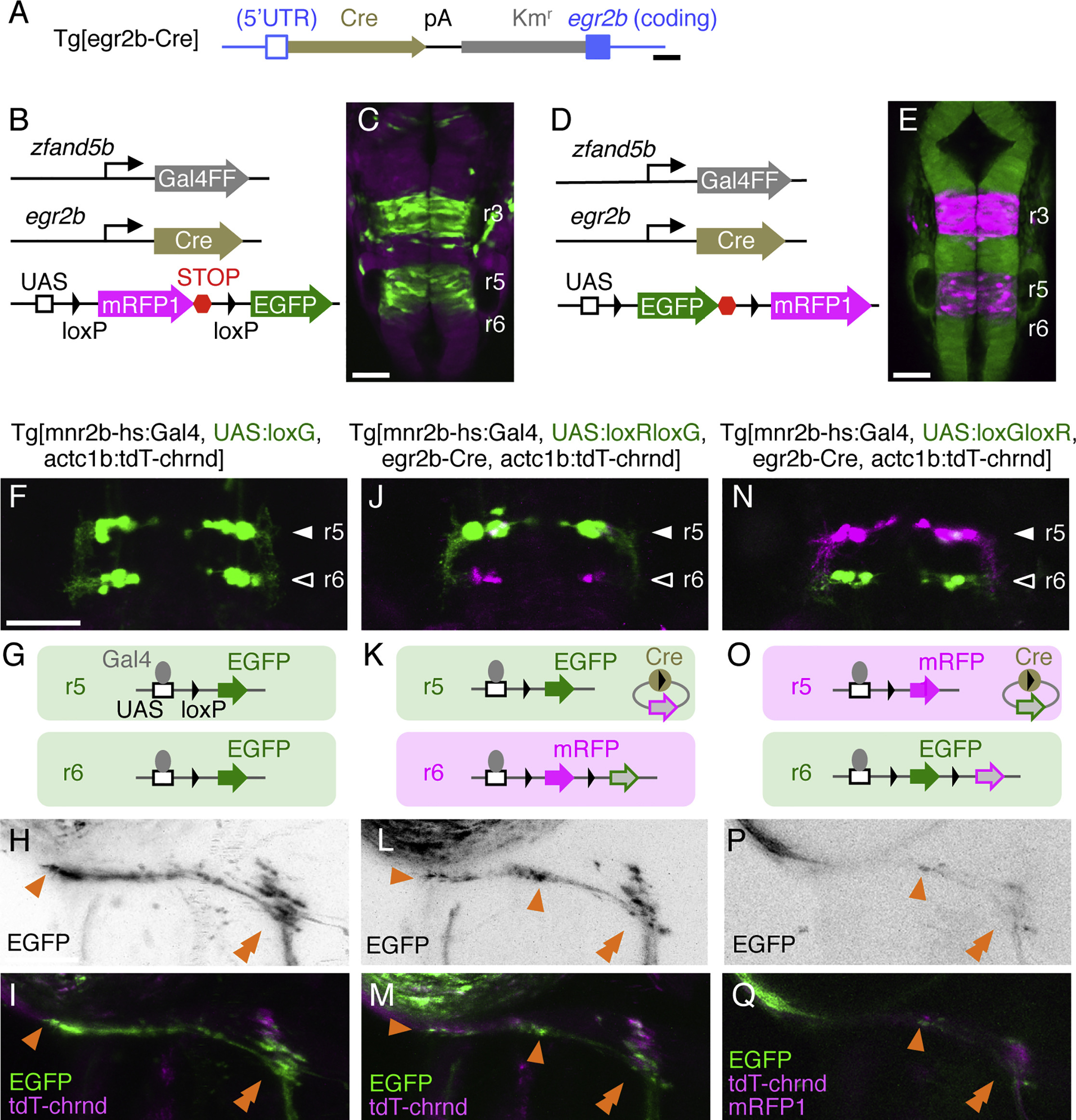
(A) Structure of Tg[egr2b-Cre]. The Cre-polyA-Kmr cassette was inserted downstream of the 5′ UTR of egr2b.
(B and C) eGFP expression in a Tg[SAGFF73A]; Tg[egr2b-Cre]; Tg[UAS:loxRloxG] embryo at 20 hpf (C). Tg[SAGFF73A] transgene ubiquitously drives Gal4 from zfand5b promoter (B) (Asakawa and Kawakami, 2009).
(D and E) EGFP expression (D) driven by the combination of Tg[SAGFF73A], Tg[egr2b-Cre], and Tg[UAS:loxGloxR] transgenes (E).
(F, J, and N) Dorsal views of r5 and r6 in larvae carrying the transgenes indicated above at 5 dpf.
(G, K, and O) Schematic illustrations of Gal4- and Cre-mediated expression of eGFP and mRFP1 in r5 and r6.
(H, I, L, M, P, and Q) Axon terminals of mnr2b-ABNs innervating the left lateral rectus muscle. Tg[UAS:loxG] is generated by spontaneous excision of the floxed mRFP1 from Tg[UAS:loxRloxG] in the germline. The orange arrowheads indicate the terminal and boutons of en grappe terminals. The double arrowheads show the CPEZ. In (Q), mRFP1 signal in mnr2b-ABNs in r6 is below detection limit.
Scale bars indicate 200 bp in (A) and 50 μm in (C), (E), (F), and (H).
|
Construct:
TgBAC(egr2b:Cre)
|