
|
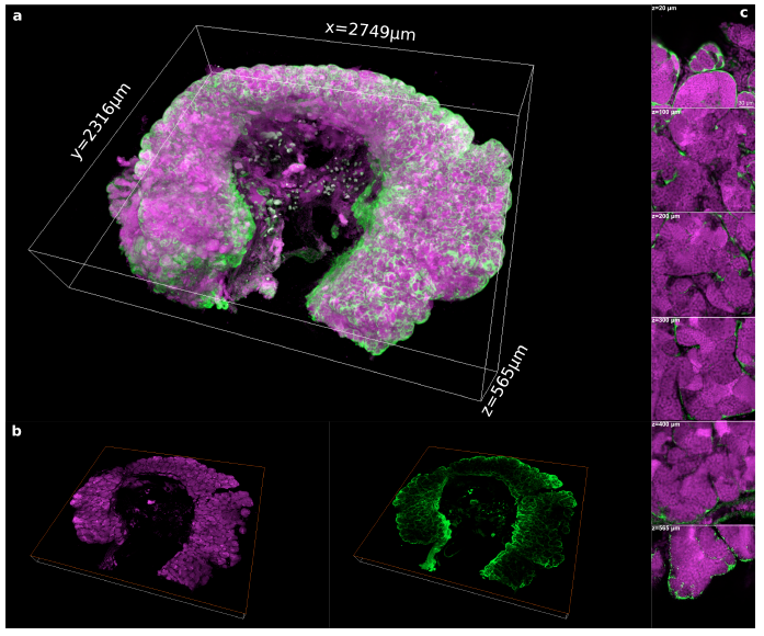
Fig. S5
3D reconstruction of a whole portion of a testis cleared by the PACT method.
(a) 3D rendering of a whole portion of a testis dissected from the transgenic line Tg(gsdf:GFP) and cleared by the PACT method. (b) 2D optical section of the whole testis at 350 μm in depth. (c) Magnified view on 2D optical sections at 20 μm, 100 μm, 200 μm, 300 μm, 400 μm and 565 μm in depth respectively. We acquired a total volume of 2.749 mm x 2.316 mm x 0.565 mm. The imaging of the whole testis took 7.5 h in our conditions and generated 28 GB of data. Images were acquired in 12 bits at a scanning speed of 600 Hz and a resolution of 512 x 512 pixels with two lines average. Voxel size: 0.865 μm x 0.865 μm x 1 μm. Nuclei are in magenta and Sertoli cells in green. Scale bar in (c) : 30 μm.