
|
Fig. S7
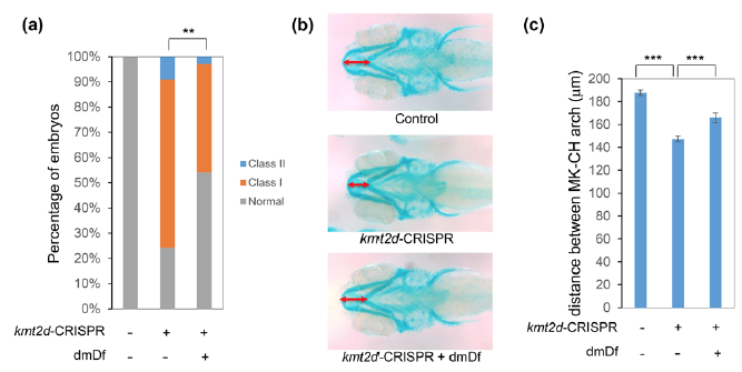
Treatment with dmDf rescues the developmental defects of kmt2d-CRISPR F0 mutants. (a) Similar to kmt2d-MO, kmt2d-CRISPR F0s also exhibit CE defects, and treatment with dmDf ameliorates the CE defects in kmt2d-CRISPR F0 embryos. (b) 5 dpf embryos were stained with Alcian blue to visualize jaw layout. kmt2d-CRISPR F0s exhibit the same change in jaw layout as kmt2d-MO-injected embryos, with a reduced distance between MK and CH cartilages. Treatment with dmDf ameliorates the jaw defects seen in kmt2d-CRISPR F0 embryos. (c) Quantitative measurement of the distance between MK and CH cartilages. ***: p< 0.001. Error bars show SEM (standard error of the mean).