
|
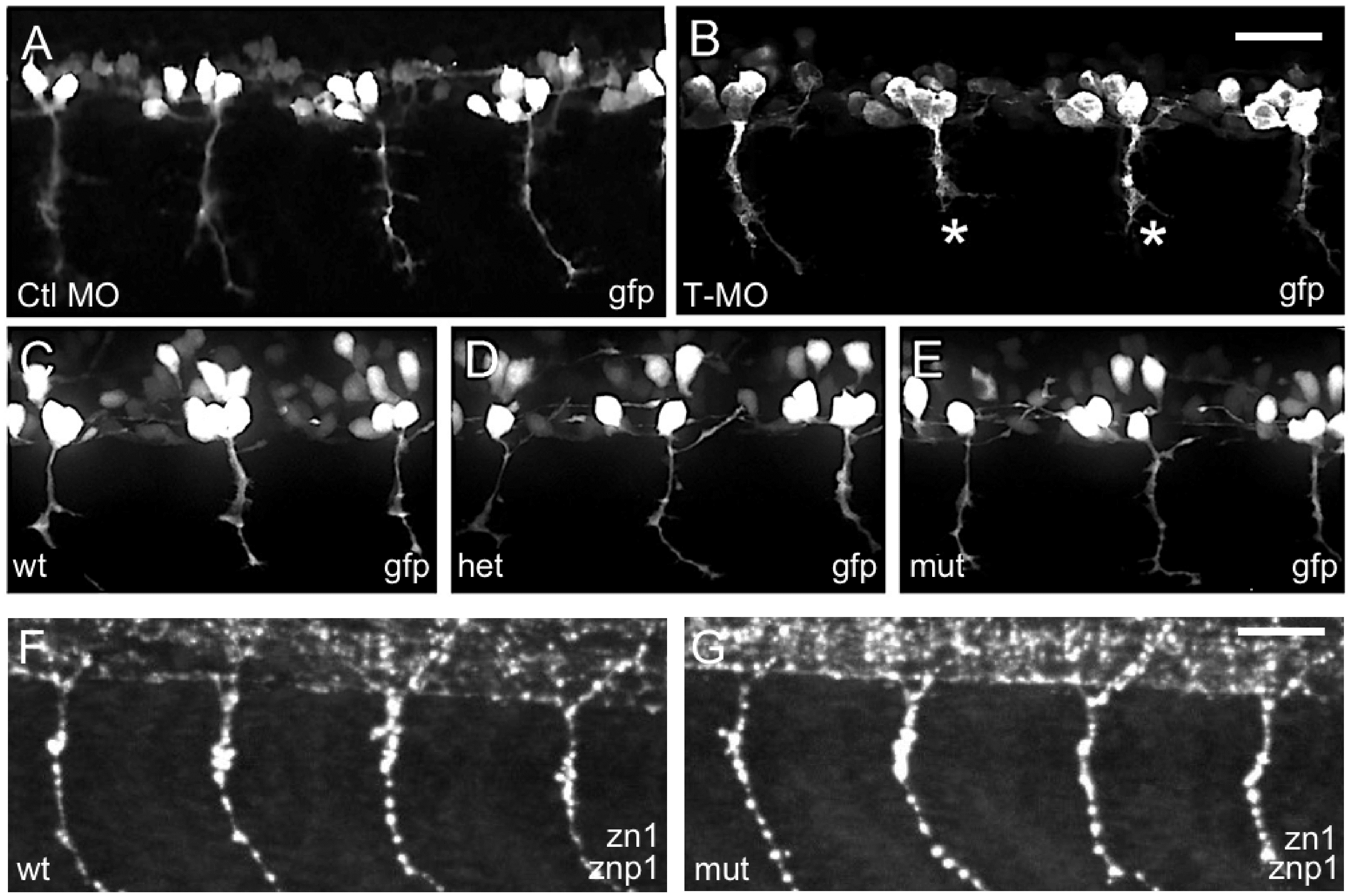
Fig. 4
Islet2a morphant but not mutant embryos displayed altered CaP morphology.
(A-E) In live tg(mnx1:gfp) 24 hpf embryos, CaP neurons expressed gfp in their somas and axons. (A, B) Injection of a T-MO (MO) led to truncation of ventrally projecting axons (asterisks, B) compared to control (Ctl, A), as previously reported [15, 17]. (C-E) In wildtype (wt, C), heterozygous (het, D) and mutant (mut, E) islet2a embryos, CaP neuron axon growth and trajectories appeared normal regardless of genotype. Sample size ranged from 8–30 per condition. Scale bar in E, for A-E: 50 μm. (F, G) In fixed non-transgenic 28 hpf embryos, zn1/znp1 immunoreactivity did not reveal any differences in CaP axon morphology between wildtype (F; n = 9) versus mutant (G; n = 9) embryos.