
Fig. 2
|
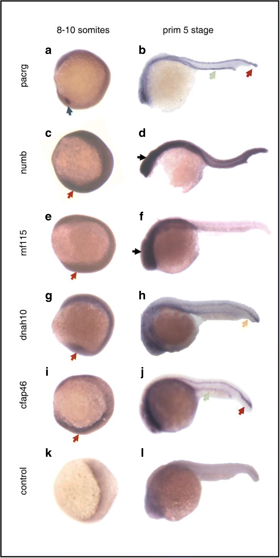
Fig. 2
Whole mount in situ hybridization analysis of candidate genes at two stages: 8–10 somites and primordium 5 stage. a, c, e, g, i, k Results of in situ hybridization of candidate genes and standard control at 13–15 hpf (8–10 somites). Embryos are viewed laterally with anterior to the top to examine KV expression. b, d, f, h, j, l Results of in situ hybridization of candidate genes and standard control at 24 hpf (primordium 5 stage). Lateral view of embryos with anterior to the left. KV (blue arrow), floor plate (red arrows), pronephric duct (green arrows), notochord (yellow arrow), head (black arrows), ubiquitous expression (orange arrows)