
|
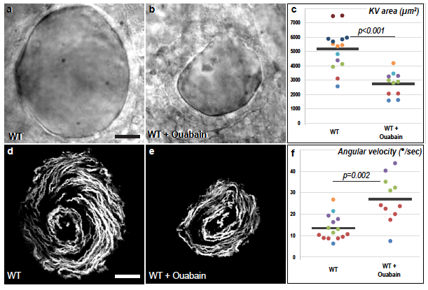
Fig. S6
The velocity of the zebrafish Left-Right Organizer flow increases upon pharmacological reduction of organ size. a-c, Ouabain treatment of WT embryos inhibits lumen inflation of Kupffer’s Vesicle (KV) and thereby reduces organ size. a,b, are brightfield images of the equatorial plane of control (a, n=15) and Ouabain-treated embryos (b, n=11). d-f, Both WT control (d) and Ouabain-treated embryos (e) display a circular flow pattern. d,e are temporal projections of the trajectories of fluorescent microspheres in the KV lumen of the embryos displayed in a and b. f, The angular velocity of the KV flow is increased upon Ouabain treatment. a,b,d,e are dorsal views of 8 somites stage KVs, anterior is up. Horizontal grey bars in c,f represent mean values. The WT control embryos displayed in this figure are also part of the dataset that is used in Fig. 4a-h. Scale bars: 20 μm.