
|
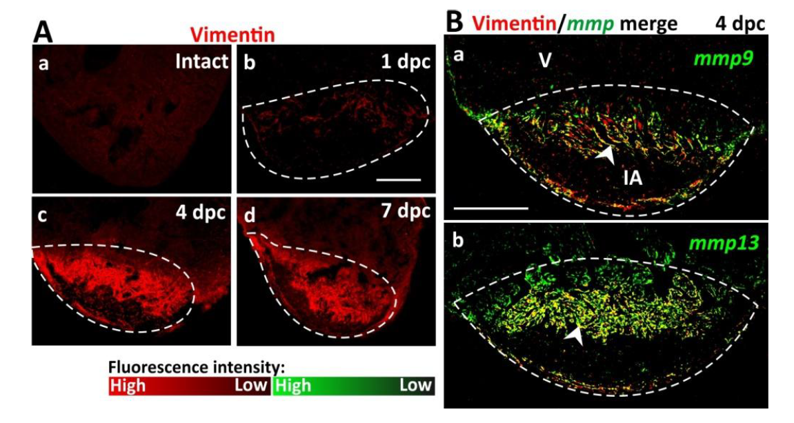
Fig. S1
Elevated numbers of vimentin-positive fibroblasts in the injured area contribute to MMP production. (A) Immunofluorescence images of cryosections to show the localization of vimentin (in red) and hence the distribution of fibroblasts in (Aa) intact (control) and (Ab-Ad) cryoinjured wild type heart ventricles (V) at: (Ab) 1 dpc, (Ac) 4 dpc, and (Ad) 7 dpc. (B) Fluorescence in situ hybridization (FISH) was conducted to show the expression of (Ba) mmp9 and (Bb) mmp13 (in green), after which immunohistochemistry was performed to show the localization of vimentin (in red) in the IA of wild type heart ventricles at 4 dpc. These representative images of paraffin sections show the FISH and immunofluorescence data when merged. Regions of co-localization appear in yellow (see white arrowheads). In each panel, the regions bounded by the dashed white lines indicate the IA. Scale bars: 200 μm.