
|
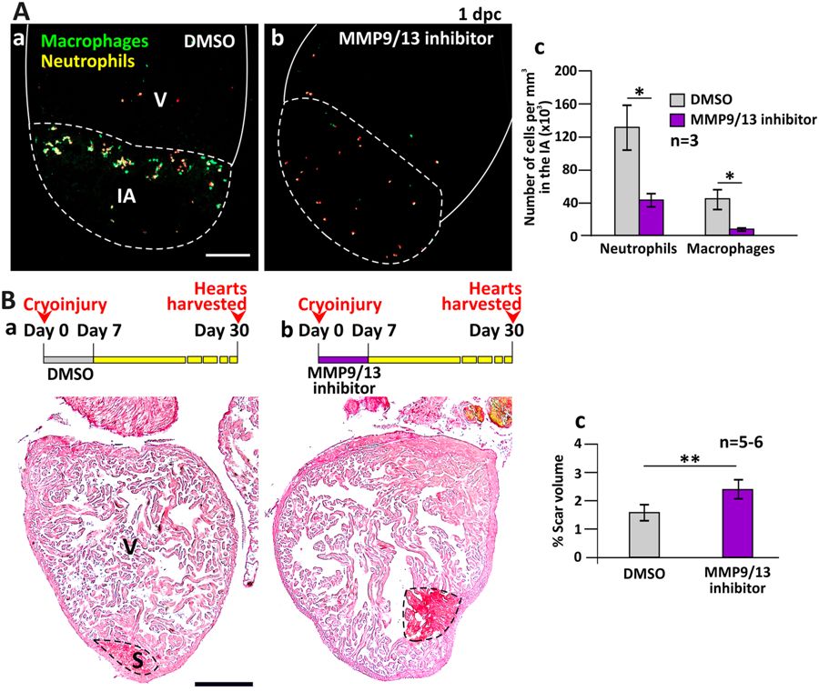
Fig. 5
MMP9/13 inhibitor I (MMP9/13 inhibitor) reduced the number of inflammatory cells into the injured area (IA) and reduced the regenerative ability of the heart. (A) Tg (coro1a:EGFP; lyz:dsRed) fish were treated with DMSO (control) or MMP9/13 inhibitor after heart ventricle cryoinjury. These images show the localization of macrophages and neutrophils in the IA of ventricles (V) at 1 dpc. Scale bar: 200 µm. (Ac) Bar graph to show the numbers of neutrophils and macrophages per mm3 in the IA of control and MMP9/13 inhibitor-treated fish at 1 dpc. (B) Representative picrosirius red stained paraffin sections of wild type fish hearts harvested at 30 dpc, after the fish were injected with: (Ba) DMSO or (Bb) MMP9/13 inhibitor, every day for the first 7 dpc. Scale bar: 200 µm. (Bc) Bar graph to show the percentage of scar volume in the ventricle following DMSO or MMP9/13 inhibitor treatment. In (Ac) and (Bc), the data are expressed as mean ± standard deviation of n = 3 and n = 5 to 6 hearts, respectively. The asterisks indicate significantly different data at p < 0.05 (*) and p < 0.01 (**), two-tailed t-test.