
|
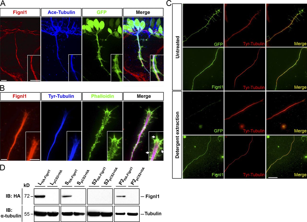
Fig. 2
Fignl1 associates with MTs in spinal motor axons. (A) Immunodetection of Fignl1 (red), acetylated α-tubulin (blue), and GFP (green) in 26-hpf Tg(Hb9:GFP) transgenic embryos. Lateral views of the trunk. (B) GC from cultured Tg(Hb9:GFP) SMN labeled with phalloidin (green), tyrosinated tubulin (blue), and Fignl1 (red) antibodies. Arrows indicate actin protrusions. (C) Detergent extraction of soluble proteins from WT or Tg(Hb9:GFP) spinal neurons immunostained by tyrosinated tubulin (red) and Fignl1 or GFP (green). Bars: (A and B) 10 µm; (C) 20 µm. (D) MT copelleting assay from COS-7 cells transfected with pCS2+-HA or pCS2+-HA-Fignl1 (HA-Fignl1). Supernatants (S) were incubated with taxol and GTP to induce MT polymerization and centrifuged on a sucrose cushion to pellet MTs and their associated proteins (P2). Soluble tubulins and proteins that did not bind MTs remained in the S2 supernatant. WBs assessed Fignl1 and α-tubulin levels in different fractions. L, total lysate.