
Fig. 3
|
Fig. 3
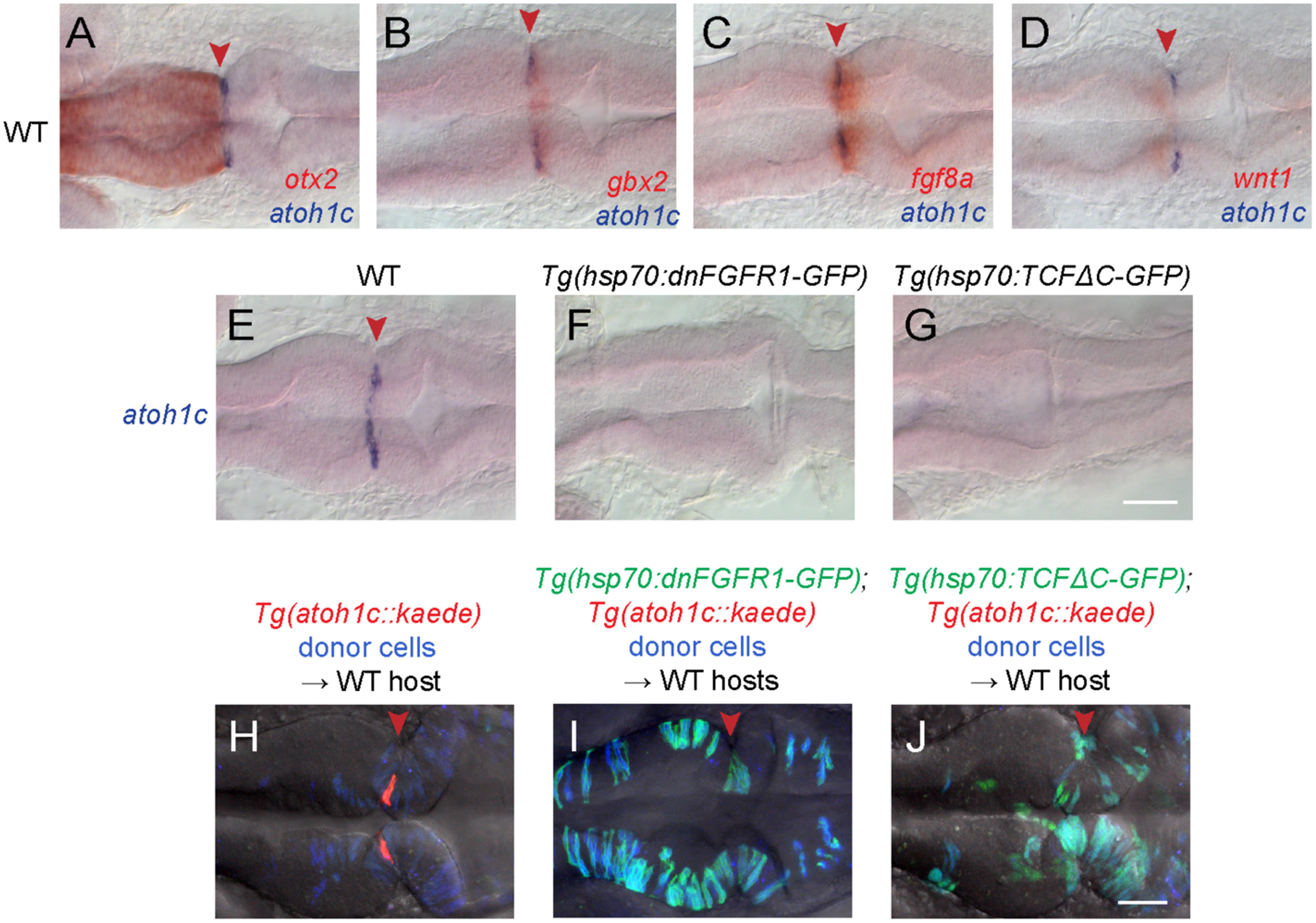
Specification of the atoh1c MHB domain. A-D: Double RNA in situs for atoh1c (blue) and otx2 (A), gbx2 (B), fgf8 (C), and wnt1 (D) (red) show that the atoh1c expression in a few cells immediately posterior to the MHB. E-G: atoh1c expression is absent when Fgf (F) or Wnt (G) signaling is blocked with heat-inducible dominant-negative transgenes activated at 10 hpf. H-J: atoh1c expression requires cell-autonomous Fgf and Wnt signaling. Live imaging of chimeras with donor-derived cells (blue) expressing dn-FGFR1 (I, green) or dn-TCFΔC (J, green) that are unable to express Tg(atoh1c::kaede) (red in the control chimera in H) even if they lie at the MHB (red arrowheads). All embryos are at 22 hpf and are shown in dorsal views with anterior to left. Scale bars: 50 µM.
Reprinted from Developmental Biology, 438(1), Kidwell, C.U., Su, C.Y., Hibi, M., Moens, C.B., Multiple zebrafish atoh1 genes specify a diversity of neuronal types in the zebrafish cerebellum, 44-56, Copyright (2018) with permission from Elsevier. Full text @ Dev. Biol.