
|
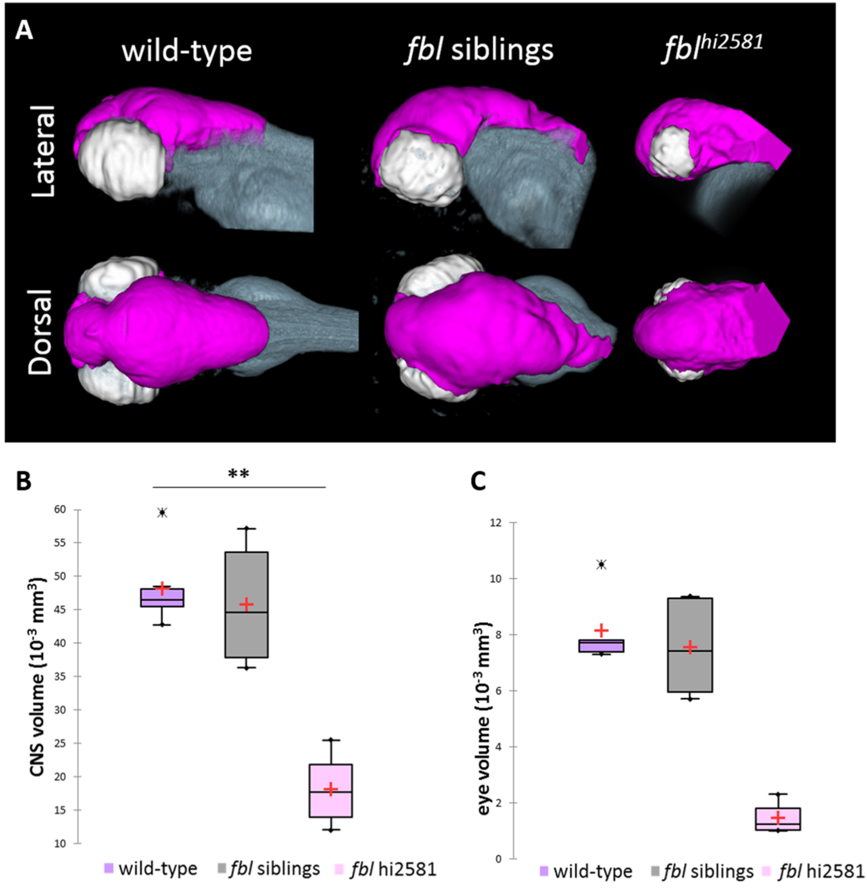
Fig. 3
The fblhi2581Tg mutation mostly affects midbrain structures from 24 hpf. (A) Volume rendering of the DiI-positive domains (gray) and surface rendering of a manual segmentation of the CNS (magenta) and eye (white) based on the DiI signal in 3dpf wild-type, fbl siblings and fblhi2581Tg mutant embryos. fbl mutant larvae display an apparent reduction of the CNS volume compared to their siblings or wild-type larvae Lateral views: anterior to the left, dorsal to the top. Dorsal view: anterior to the left, right to the top. (B) Quantification of mean CNS volume highlights a significant difference between fbl mutants, their siblings and wild-type larvae. Statistical analyses were performed on six samples per condition. p-value: .003 (Kruskal-Wallis test). (C) Quantification of mean eye volume highlights a significant difference between fbl mutants, their siblings and wild-type larvae. Statistical analyses were performed on the mean size of both eye of six samples per condition. p-value: .005 (Kruskal-Wallis test). Purple: wild-type, gray: fbl siblings, pink: fblhi2581Tg. Scale bar: 100 µm. Anterior is to the left.
Reprinted from Developmental Biology, 437(1), Bouffard, S., Dambroise, E., Brombin, A., Lempereur, S., Hatin, I., Simion, M., Corre, R., Bourrat, F., Joly, J.S., Jamen, F., Fibrillarin is essential for S-phase progression and neuronal differentiation in zebrafish dorsal midbrain and retina, 1-16, Copyright (2018) with permission from Elsevier. Full text @ Dev. Biol.