
|
Fig. S3
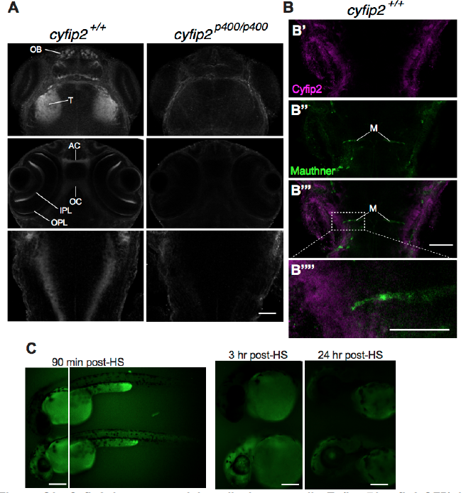
Cyfip2 is expressed broadly in neuropil; Tg(hsp70:cyfip2-GFP) is detectable in CNS for approximately 24 hours after heat shock, Related to Figure 2 (A) Cyfip2 Ab stain in cyfip2+/+ and cyfip2p400/p400 larvae at 72 hpf. OB: olfactory bulb, T: tectum, AC: anterior commissure, OC: optic chiasm, IPL: inner plexiform layer, OPL: outer plexiform layer (scale bar 30 μm). (B) Cyfip2 Ab stain (magenta) at 72 hpf showing labeling near the lateral dendrite of the Mauthner cell (M, green), labeled by Tg(GFFDMC130A);Tg(UAS:gap43-citrine) (scale bar: B’-B’’’: 30 μm; B’’’’: 15 μm). (C) Expression of Tg(hsp70:cyfip2-GFP) after a single 40 min heat shock at 37°C in larvae with (bottom) and without (top) the transgene. Scale bars 100 μm.