
Fig. 2
|
Fig. 2
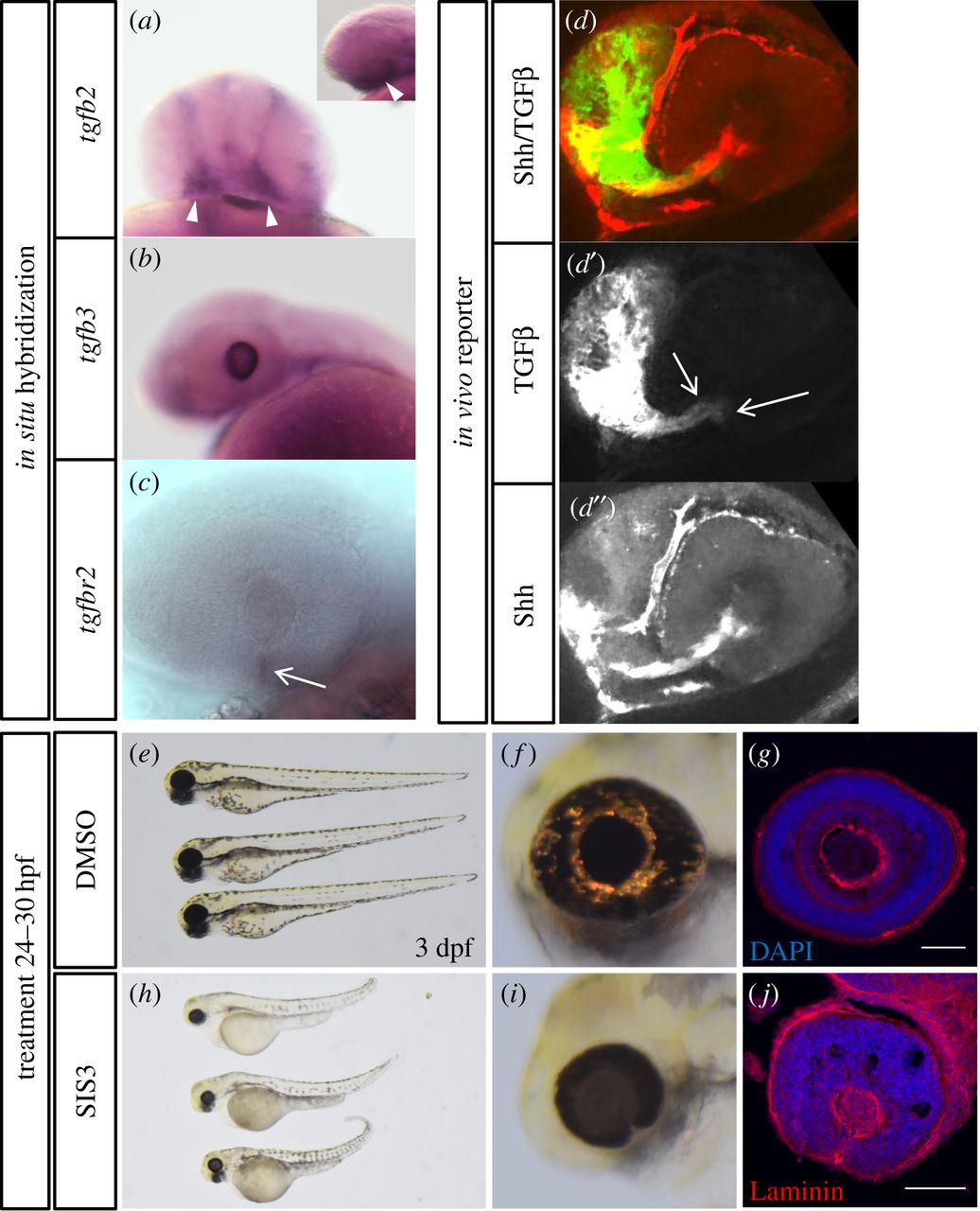
TGFβ in the zebrafish eye. (a) Whole-mount in situ hybridization (WMISH) of tgfb2 (30 hpf), frontal view. Small image shows a lateral view. Note expression in periocular tissue (arrowheads). (b) WMISH of tgfb3 (30 hpf), lateral view. Note expression in the developing lens. (c) WMISH of tgfbr2 (30 hpf), lateral view. Note expression in the optic fissure (arrow). (d) In vivo signalling for TGFβ (green) and Shh (red) for orientation, split into TGFβ (d′) and Shh (d″), at 24 hpf. Active TGFβ signalling in the optic fissure margins (d′, arrows). (e–j) Embryos (3 dpf) treated with DMSO (e–g) or specific inhibitor of Smad3 (SIS3, h–j) from 24 to 30 hpf. Sagittal sections through eyes of (g) DMSO and (j) SIS3-treated embryos at 3 dpf, stained with DAPI and anti-Laminin. SIS3-treated embryos show a persisting optic fissure with persisting basal lamina and absence of retinal layering.