
|
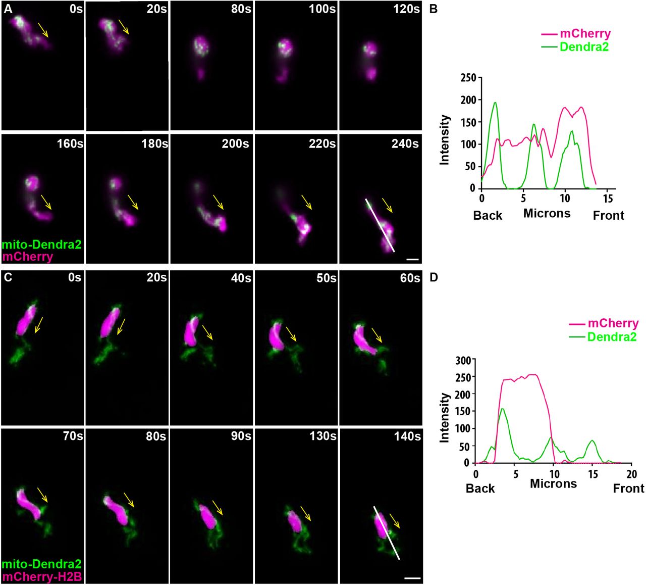
Fig. 1
Mitochondria localize to both the front and the rear of neutrophils. (A) Random motility of neutrophils in the head mesenchyme of a 3 dpf larvae of Tg(lyzC:mitoDendra2)p14 crossed with Tg(lyzC:mCherry). (B) Quantification of fluorescence intensity along the indicated line (at 240 s). (C) Tg(lyzC:mitoDendra2)pu14 was crossed with Tg(lyzC:mCherry-H2B) to visualize mitochondria network relative to the nucleus. (D) Quantification of fluorescence intensity along the indicated line (at 140 s). Montages of one representative movie out of eight are shown. Arrows indicate the direction of cell migration. Scale bars: 5 µm.