
Image
|
Figure Caption
Fig. 3
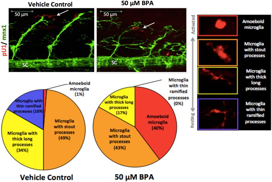
Activated microglia associate with defective motor neurons. Double transgenic Tg:mnx1-GFP/pU1-RFP embryos identify microglia (red) and motor neurons (green). White arrows point to pU1+ microglial cells spatially associated with motor neurons at 48 hpf. Percent values in the pie charts show the relative proportion of total microglia in each activation state compared to total number of microglia cells in region of interest (right panel) when exposed to BPA compared to vehicle controls (N = 9–12 biological replicates; N = 1–10 technical replicates). SC = spinal cord.
Figure Data
Acknowledgments
This image is the copyrighted work of the attributed author or publisher, and
ZFIN has permission only to display this image to its users.
Additional permissions should be obtained from the applicable author or publisher of the image.
Full text @ Sci. Rep.