
Image
|
Figure Caption
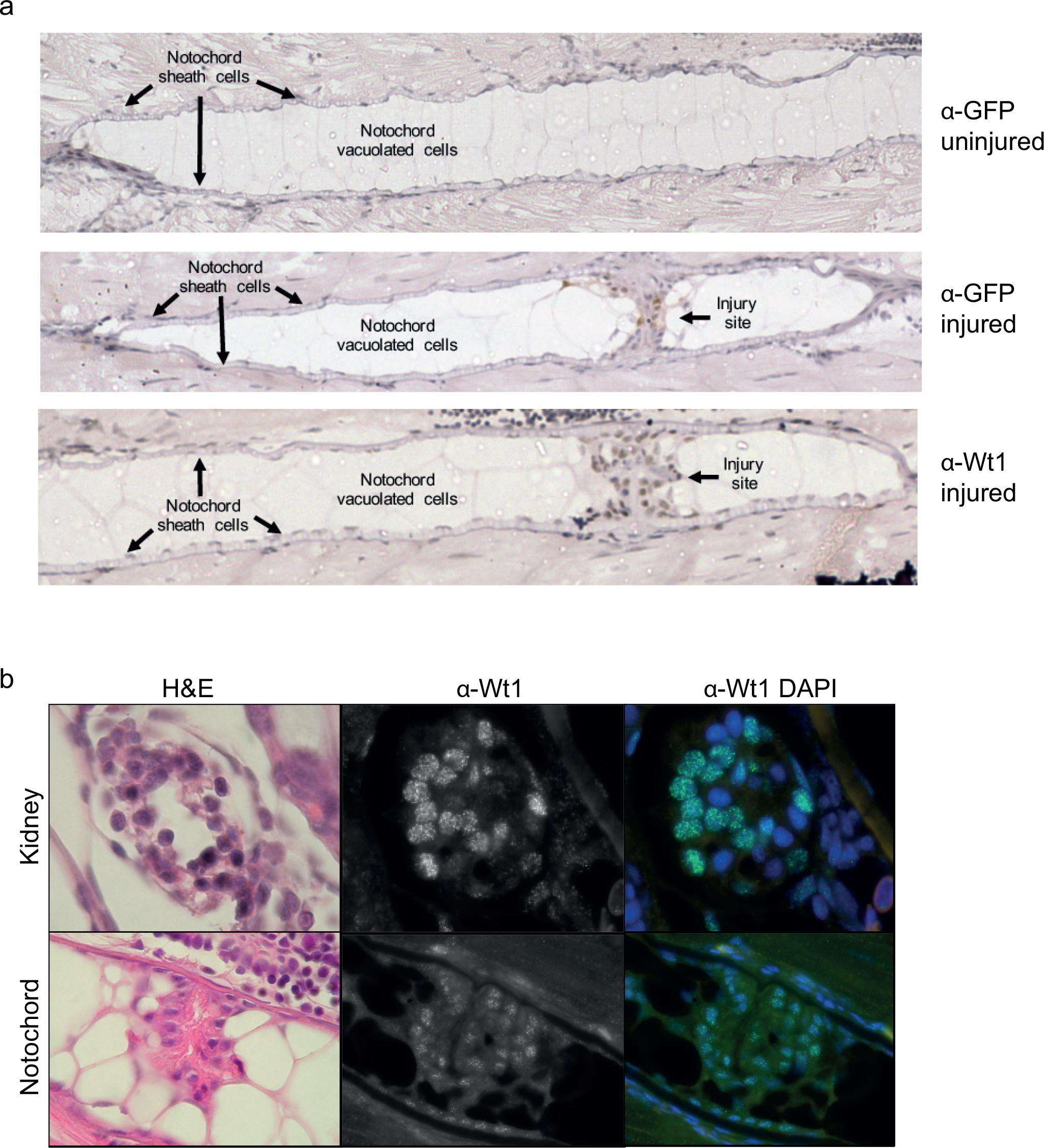
Fig. 1-S2
Wt1 and GFP protein expression in the notochord.
(a) Immunohistochemistry of injured and uninjured notochord regions, stained with α-GFP and α-Wt1 antibodies. Sheath cells and vacuolated cell populations, and injury site are indicated. n > 10; five experimental replicates. (b) Immunofluorescence of 3 dpi injured notochord tissue stained with α-Wt1 antibody. Kidney tissue is shown as a positive control. dpi: days post injury. Experimental replicates = 5; n = 5.
Figure Data
Acknowledgments
This image is the copyrighted work of the attributed author or publisher, and
ZFIN has permission only to display this image to its users.
Additional permissions should be obtained from the applicable author or publisher of the image.
Full text @ Elife