
|
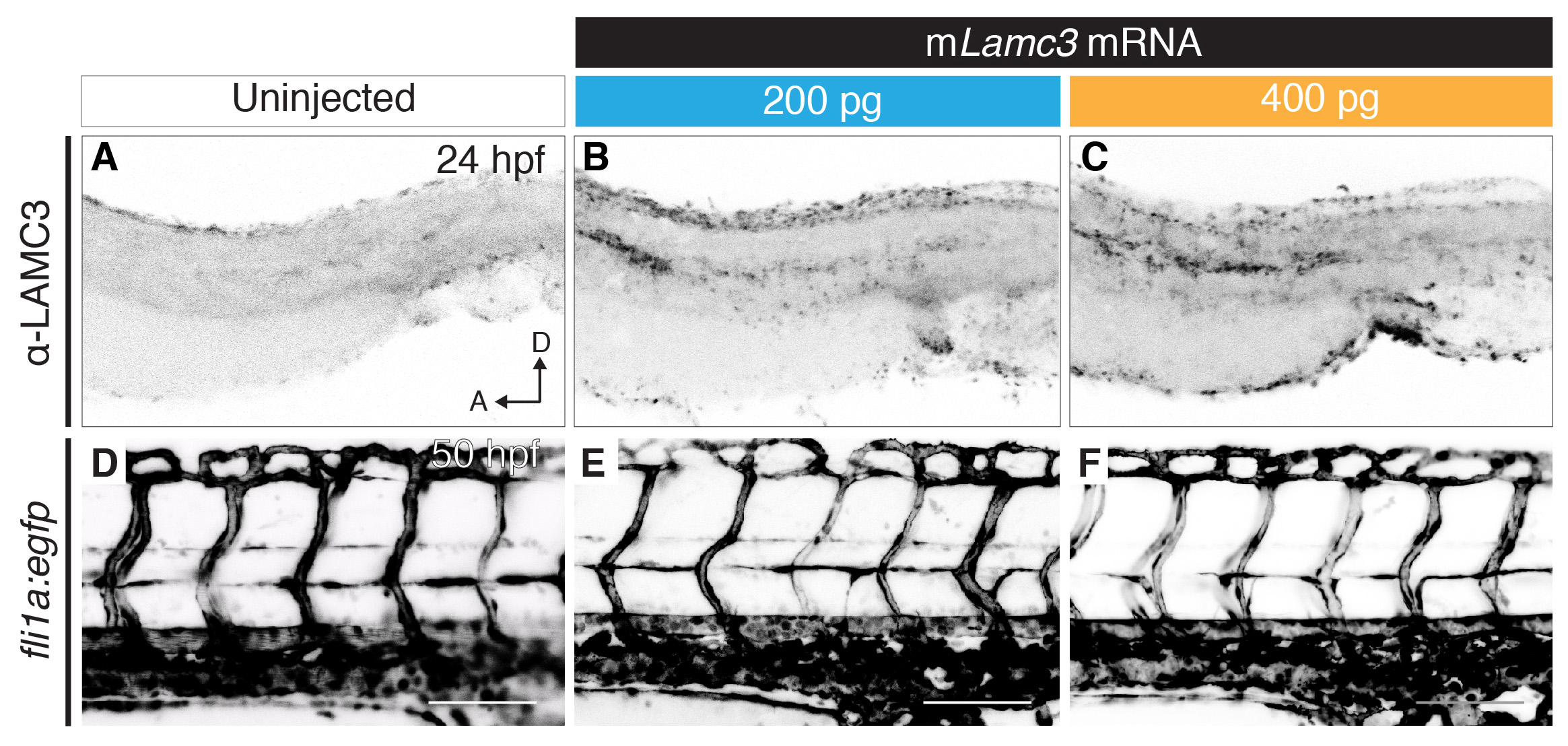
Fig. S2
Overexpression of mouse Lamc3 mRNA. (A–C) Anti-LAMC3 immunostaining in the dissected trunks of 24 hpf embryos either uninjected (A), injected with 1 nl of 200 ng/μl of mouse Lamc3 (mLamc3) mRNA (B) or injected with 1 nl of 400 ng/μl of mouse Lamc3 mRNA shows increase of antibody staining at the horizontal myoseptum, dorsal plate of the neural tube and cloaca. (D–F) Imaging of the trunk vasculature in Tg(fli1a:egfp) 50 hpf embryos either uninjected (D), injected with 1 nl of 200 ng/μl of mouse Lamc3 mRNA (E) or injected with 1 nl of 400 ng/μl of mouse Lamc3 mRNA (F). A, anterior; D, dorsal; hpf, hours post fertilisation. Scale bars: 100 μm.