
|
Fig. S17
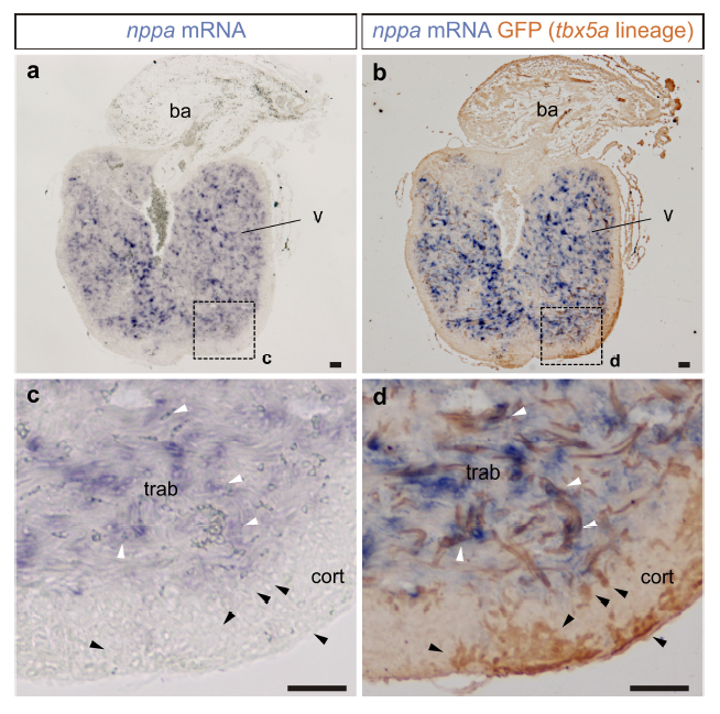
tbx5a-derived cardiomyocytes within the cortical layer are nppa negative.
nppa mRNA in situ hybridisation (a) on sagittal sections of tbx5a:mCherryp-2a-CreERT2;ubb:loxP-lacZ-loxP-GFP double transgenic animals, recombined before injury and fixed at 90 days postinjury (dpi) followed by GFP immunofluorescence (b) nppa is expressed in the trabecular layer (white arrowheads). In the cortical region, tbx5a-derived GFP+ cells are visible, which are not positive for the trabecular marker nppa (black arrowheads, n=7/7). c and d are a zoomed with of boxed areas in a and b. ba, bulbus arteriosus; cort, cortical layer; trab, trabecular layer; v, ventricle. Scale bars, 50 μm