
|
Fig. S2
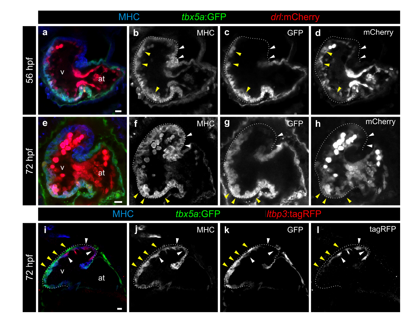
The pattern of tbx5a:GFP expression is similar to that of drl:mCherry and complementary to ltbp3:mCherry. a–h Confocal optical sections of 56 (n=5/5) and 72 (n=7/7) hours postfertilisation (hpf) tbx5a:GFP;drl:mCherry double transgenic zebrafish larvae. GFP (green) labels tbx5a+ cells, mCherry (red) drl+ cells and anti-Myosin Heavy Chain (MHC) immunofluorescence labels all cardiomyocytes. The ventricle is outlined with dotted lines. The tbx5a:GFP- and drl:mCherry- distal ventricle is marked with white arrowheads, while the yellow arrowheads point to the domains positive for both markers. Note that drl:mCherry is also expressed in endocardial cells and red blood cells in the lumen of the heart. i–l Confocal optical sections of 72 hpf hearts from tbx5a:GFP embryos injected with ltbp3:TagRFP-2A-Cre at the one cell stage. Shown is a representative heart out of 8 larvae. GFP labels tbx5a+ cells, mCherry ltbp3+ cells, and MHC all cardiomyocytes. The yellow arrowheads point to tbx5a:GFP+ cells that are ltbp3:mCherry- while the white arrowhead points to a ltbp3:mCherry+ cells within the tbx5a:GFP- domain. at, atrium; v, ventricle. Scale bars, 10 μm