
Image
|
Figure Caption
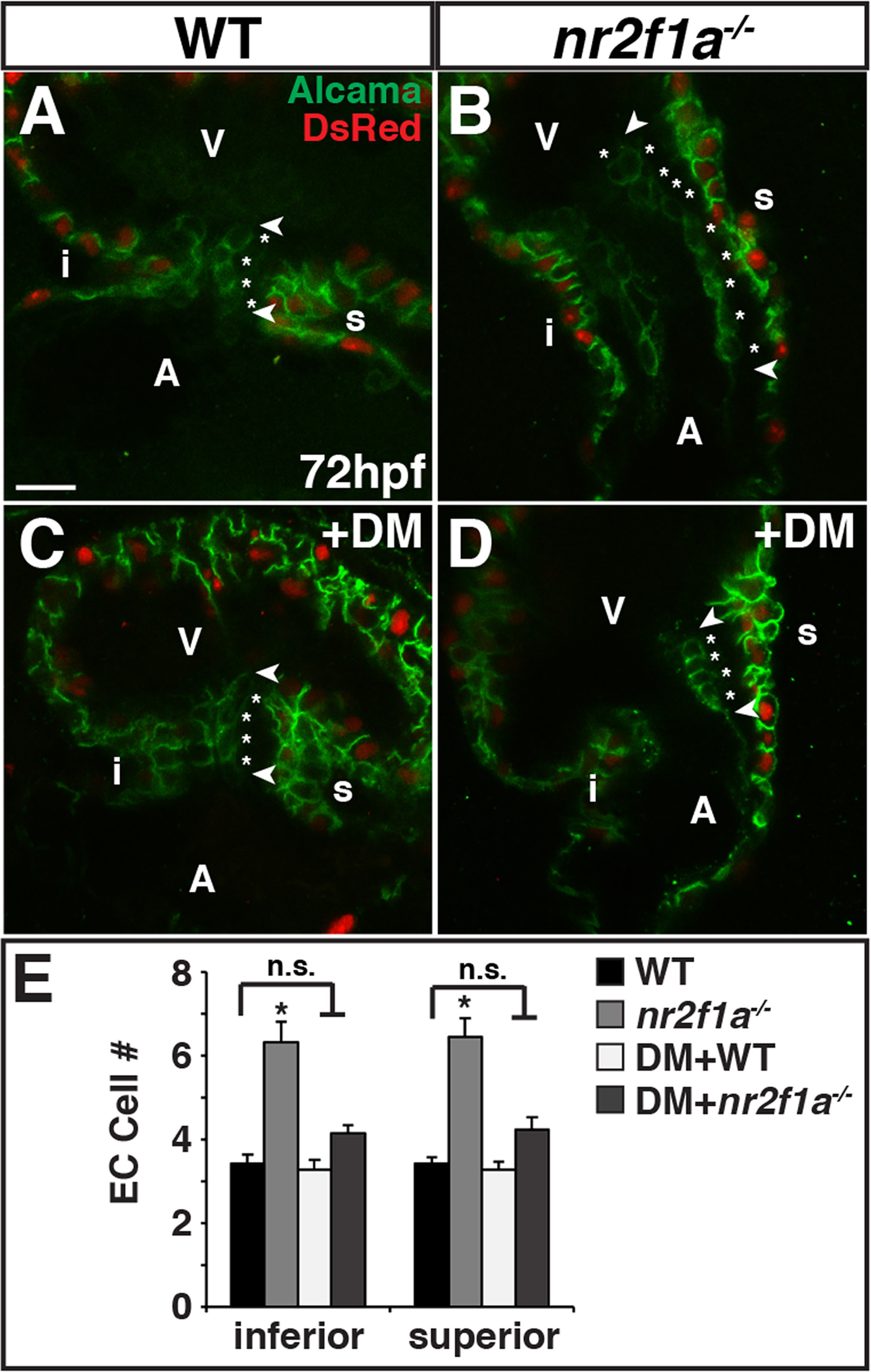
Fig. 4
Nr2f1a functions upstream of Bmp signaling to restrict ECs. (A-D) IHC of hearts from myl7: DsRed-NLS WT, nr2f1a mutant, WT DM-treated, and nr2f1a mutant DM-treated embryos for Alcama (green) and DsRed (myocardial nuclei – red). Images are frontal views of confocal stacks. Length of ECs (arrowheads). Individual EC cells (asterisks). Scale bar indicates 10 µm in A-D. (E) Mean EC number from inferior and superior valve sides in WT (n = 12), nr2f1a mutant (n = 9), WT DM-treated (n = 11), and nr2f1a mutant DM-treated (n = 13) embryos.
Figure Data
Acknowledgments
This image is the copyrighted work of the attributed author or publisher, and
ZFIN has permission only to display this image to its users.
Additional permissions should be obtained from the applicable author or publisher of the image.
Reprinted from Developmental Biology, 434(1), Duong, T.B., Ravisankar, P., Song, Y.C., Gafranek, J.T., Rydeen, A.B., Dohn, T.E., Barske, L.A., Crump, J.G., Waxman, J.S., Nr2f1a balances atrial chamber and atrioventricular canal size via BMP signaling-independent and -dependent mechanisms, 7-14, Copyright (2017) with permission from Elsevier. Full text @ Dev. Biol.