
IMAGE
Fig. S9
- ID
- ZDB-IMAGE-180516-26
- Publication
- Ojeda Naharros et al., 2017 - Loss-of-function of the ciliopathy protein Cc2d2a disorganizes the vesicle fusion machinery at the periciliary membrane and indirectly affects Rab8-trafficking in zebrafish photoreceptors
- All Figures
- Figures for Ojeda Naharros et al., 2017
Image
|
Figure Caption
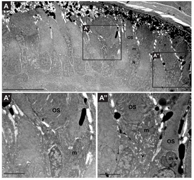
Fig. S9
Retinae of rhod:mCherry-rab8a are structurally healthy.
(A) Transmission electron microscopy image of a wild-type 5dpf tg(rhod:mCherry-rab8a) retina. The retinal structure remains normal and extension of outer segments is unaffected. Variable accumulation of membrane-bound structures in inner segment regions is observed in a subset of PRs as a consequence of the overexpression of the transgenic construct (absent in A’, present in A”). Scale bars: 10 μm in A and 2 μm in A’ and A”. OS outer segment, m mitochondria, N nucleus, v vesiculo-tubular structures.
Acknowledgments
This image is the copyrighted work of the attributed author or publisher, and
ZFIN has permission only to display this image to its users.
Additional permissions should be obtained from the applicable author or publisher of the image.
Full text @ PLoS Genet.