
Fig. 9
- ID
- ZDB-IMAGE-180516-19
- Antibodies
- Publication
- Ojeda Naharros et al., 2017 - Loss-of-function of the ciliopathy protein Cc2d2a disorganizes the vesicle fusion machinery at the periciliary membrane and indirectly affects Rab8-trafficking in zebrafish photoreceptors
- All Figures
- Figures for Ojeda Naharros et al., 2017
|
Fig. 9
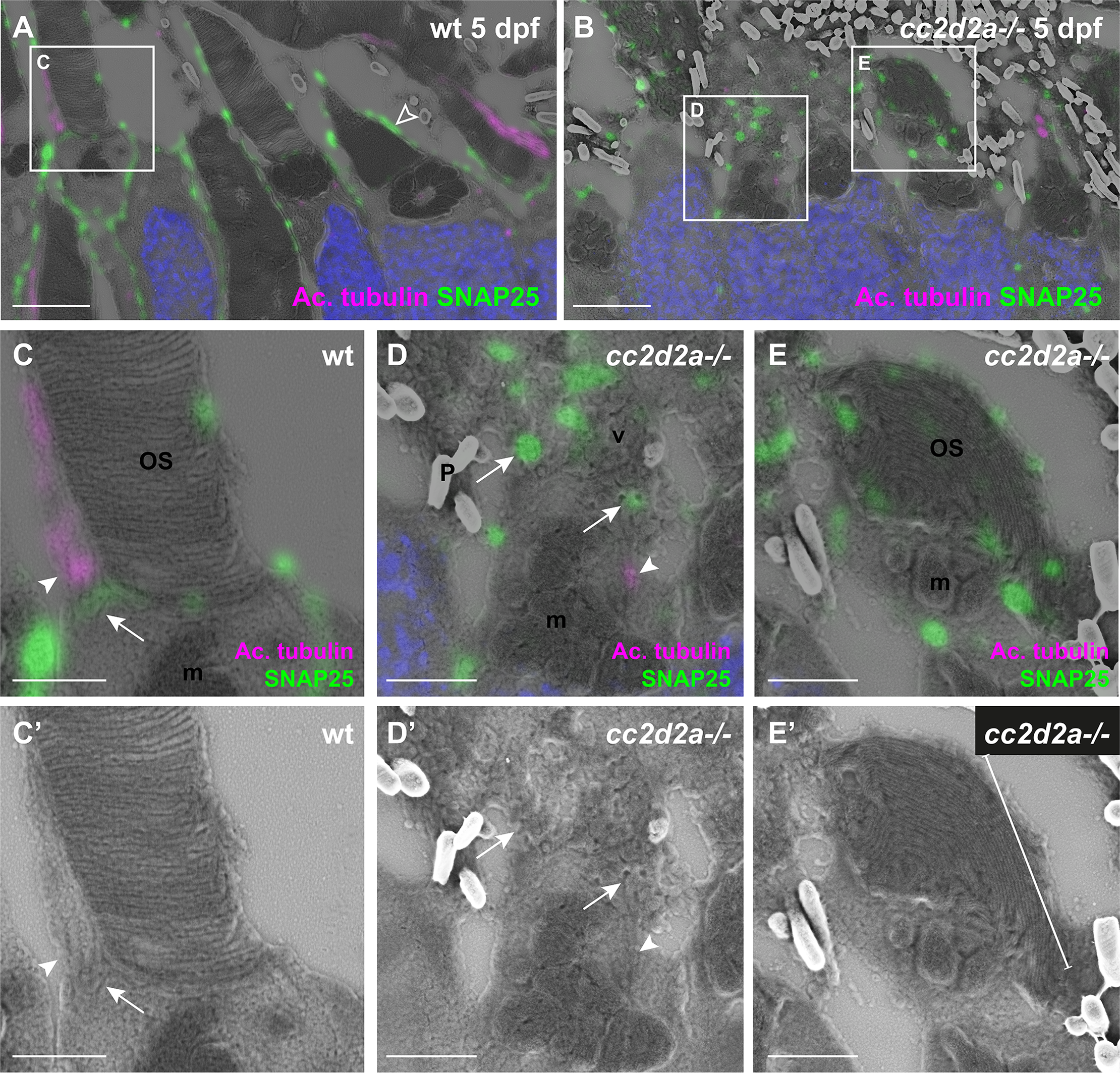
SNAP25 localizes to wt periciliary membrane and mislocalizes to accumulated vesicles of cc2d2a-/- PRs (A-E) 5 dpf CLEM retinal sections stained for SNAP25 (green), acetylated tubulin (magenta) and DAPI (blue, nuclei). (C-E) are higher magnification images of the boxed areas in (A, B) and (C’-E’) are the corresponding SEM images. In wt PRs (A) SNAP25 is found at the inner segment apical membrane, along the calycal processes (empty arrowhead in A) as well as at the periciliary membrane (C-C’, arrow) around the base of the anti-acetylated tubulin-marked cilium (C-C’, arrowhead). In contrast, in cc2d2a-/- PRs (B), SNAP25 prominently mislocalizes in accumulated vesicles (D-D’, arrows) and dysmorphic OSs (E-E’, bracket). Arrowheads point to connecting cilia in (C-D’). Scale bars: 4 μm in A-B, 1 μm in C-E’. OS outer segment, m mitochondria, N nuclei, P pigment, v vesicles, wt wild-type.