
Image
|
Figure Caption
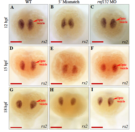
Fig. S4
WISH analysis of WT and rnf152 MO with rx2- specific probe identified alteriations in the optic vesicles at 12,15, and 18 hpf.
(A-C) 12 hpf, (D-F) 15 hpf, and (G-I) 18 hpf. (A,D,&G) WT embryos, (B,E,&H) Embryos injected with 5' mismatch control morpholino, and (C,F,&I) Embryos injected with rnf152 morpholino. Level of rx2 transcripts did not show significant differences in the optic vesicles among WT, 5' mismatch control, and rnf152 MO at 12, 15, and 18 hpf. Red arrowheads indicate optic vesicle of the embryo (n=3). Scale Bars A-I: 50 μm.
Figure Data
Acknowledgments
This image is the copyrighted work of the attributed author or publisher, and
ZFIN has permission only to display this image to its users.
Additional permissions should be obtained from the applicable author or publisher of the image.
Full text @ Mol. Cells