
Image
|
Figure Caption
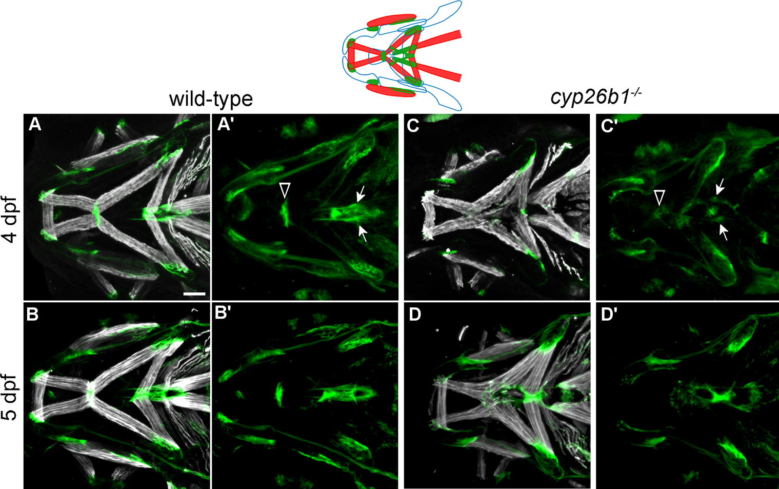
Fig. 5 Loss of Cyp26b1 function disrupts cranial tendon differentiation. (A-B) Tsp4b (green) is enriched at the muscle attachment sites in wild-type zebrafish at 4 and 5 dpf. (C) At 4 dpf, cyp26b1 mutants display Tsp4b at jaw muscle attachments, though weakly at the mandibulohyoid junction (arrowheads in A’,C’) and sternohyoideus tendons (arrows in A’,C’). (D) At 5 dpf, punctate deposits of Tsp4b can be seen at all ectopic points of jaw muscle attachment. All images ventral view, anterior to the left. Scale bar = 50 μm.
Figure Data
Acknowledgments
This image is the copyrighted work of the attributed author or publisher, and
ZFIN has permission only to display this image to its users.
Additional permissions should be obtained from the applicable author or publisher of the image.
Full text @ PLoS Genet.