
Image
|
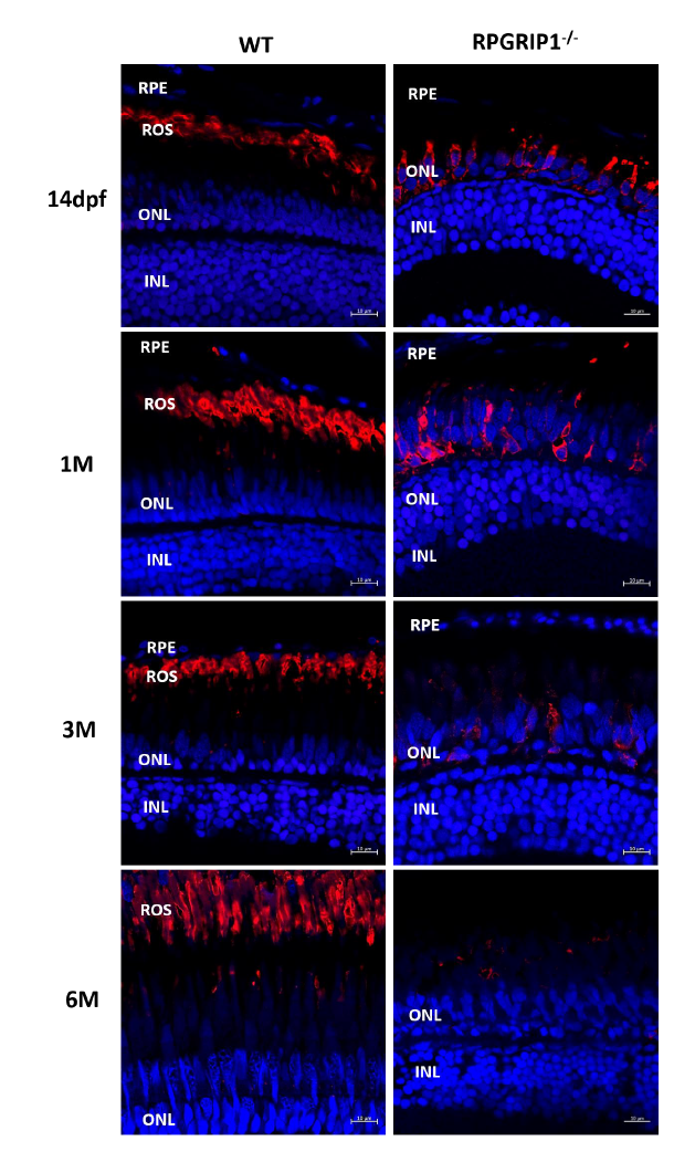
Figure Caption
Fig. S5
Immunostaining of retinal sections from wildtype and rpgrip1-/- zebrafish at age of 14 days (dpf), one month (1 mpf), 3 months (3 mpf) and 6 months (6 mpf). 4D2, labelling rhodopsin, was used for the staining. Nuclei were shown in blue using DAPI. Rhodopsin was mislocalized in all examined rpgrip1-/- mutant retina. The signal of rhodopsin was significantly decreased at 1mpf, almost absent at 3mpf, and at background level at 6 mpf. INL, inner nuclear layer; ONL, outer nuclear layer; ROS, rod outer segment; RPE, retinal pigment epithelium
Acknowledgments
This image is the copyrighted work of the attributed author or publisher, and
ZFIN has permission only to display this image to its users.
Additional permissions should be obtained from the applicable author or publisher of the image.
Full text @ Sci. Rep.