
Image
|
Figure Caption
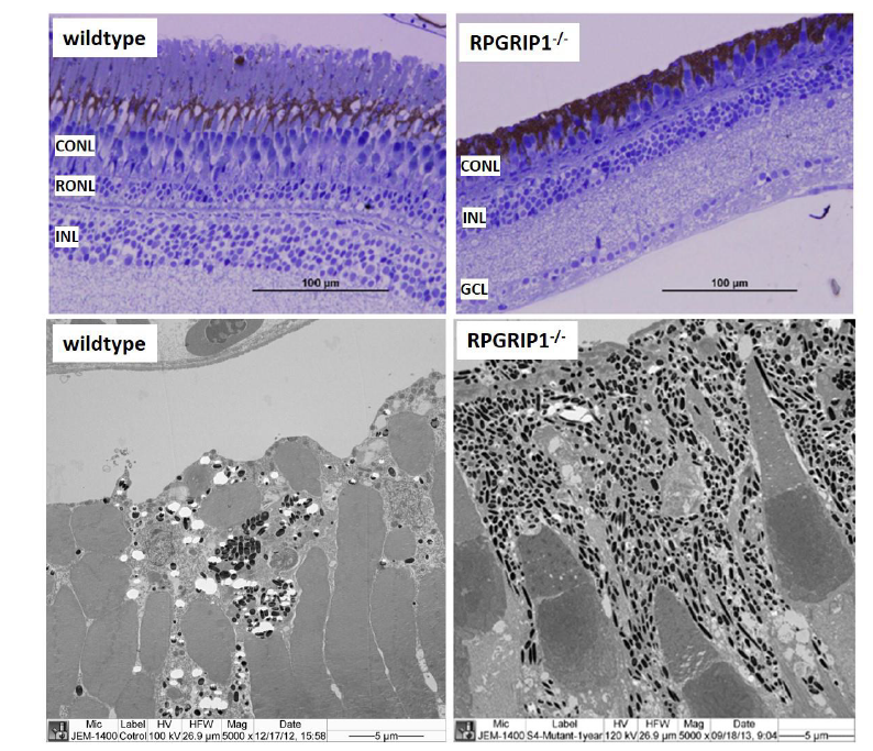
Fig. S4
Retinal structure of wildtype and rpgrip1-/- zebrafish at age of 13 months showed only short cones remained in rpgrip1-/- zebrafish retina. Upper panel, light microscopy structure of wildtype and rpgrip1-/- zebrafish retinas; lower panel, ultrastructure of wildtype and rpgrip1-/- zebrafish retina. CONL, cone outer nuclear layer; GCL, ganglion cell layer; INL, inner nuclear layer; RONL, rod outer nuclear layer.
Acknowledgments
This image is the copyrighted work of the attributed author or publisher, and
ZFIN has permission only to display this image to its users.
Additional permissions should be obtained from the applicable author or publisher of the image.
Full text @ Sci. Rep.