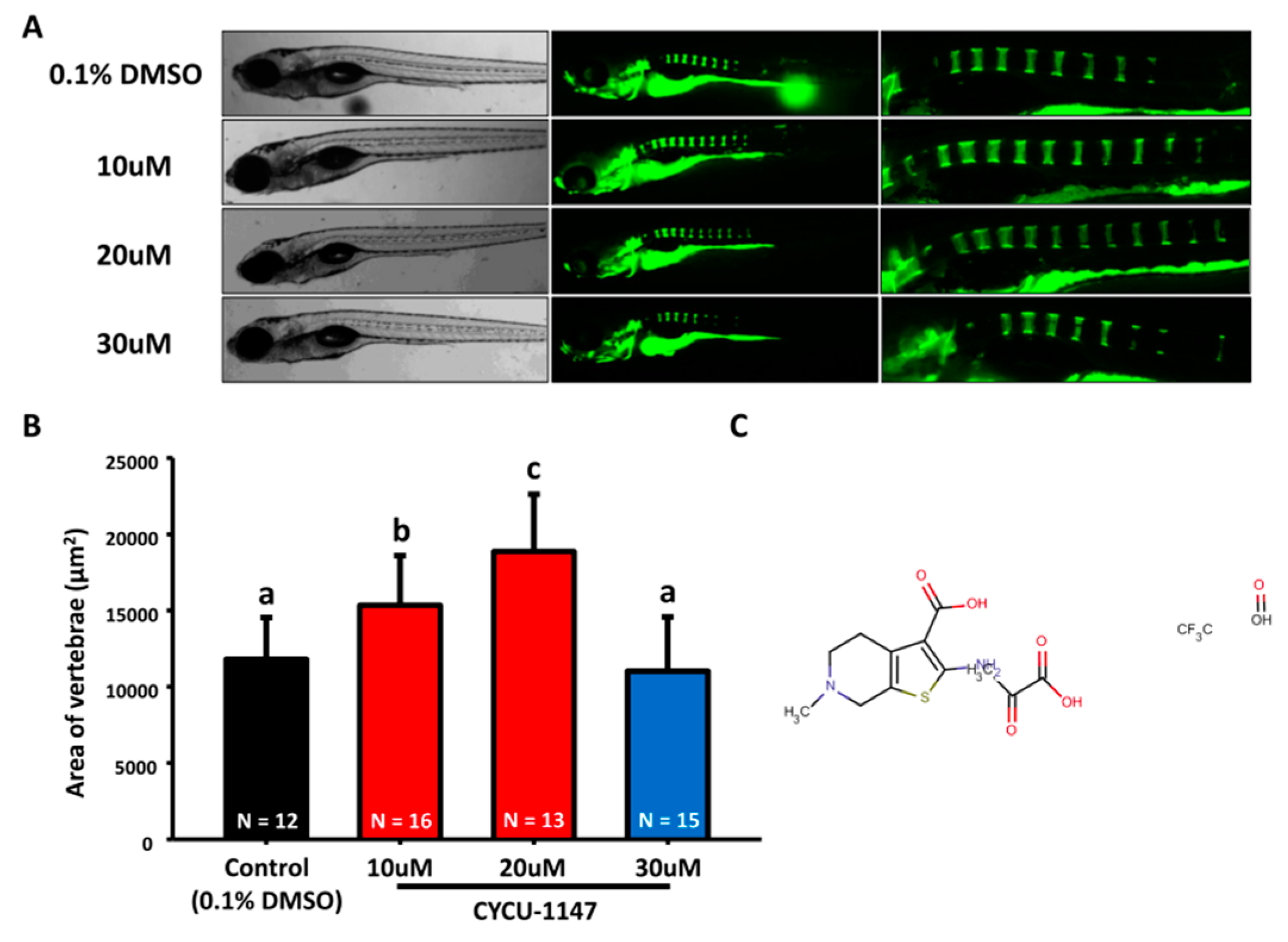
Fig. 5 Increase of mineralization in BML-267-treated zebrafish. (A) The gross morphology of zebrafish aged at 7 dpf which have been treated with different concentrations of BML-267 (10, 20, and 30 µM, left bright-filed panel) from 3 dpf onwards. Calcein staining on control and BML-267-treated embryos at 7 dpf (right green fluorescent panel); (B) Quantification of mineralization degree detecting the fluorescence intensity at the area of centrum form ring in the notochord. (values are mean ± SD; tested by one-way ANOVA pairwise comparison; N = fish number; Different letters indicate significant differences) (C) Chemical structure of BML-267.
Image
Figure Caption
Acknowledgments
This image is the copyrighted work of the attributed author or publisher, and
ZFIN has permission only to display this image to its users.
Additional permissions should be obtained from the applicable author or publisher of the image.
Full text @ Molecules

