
|
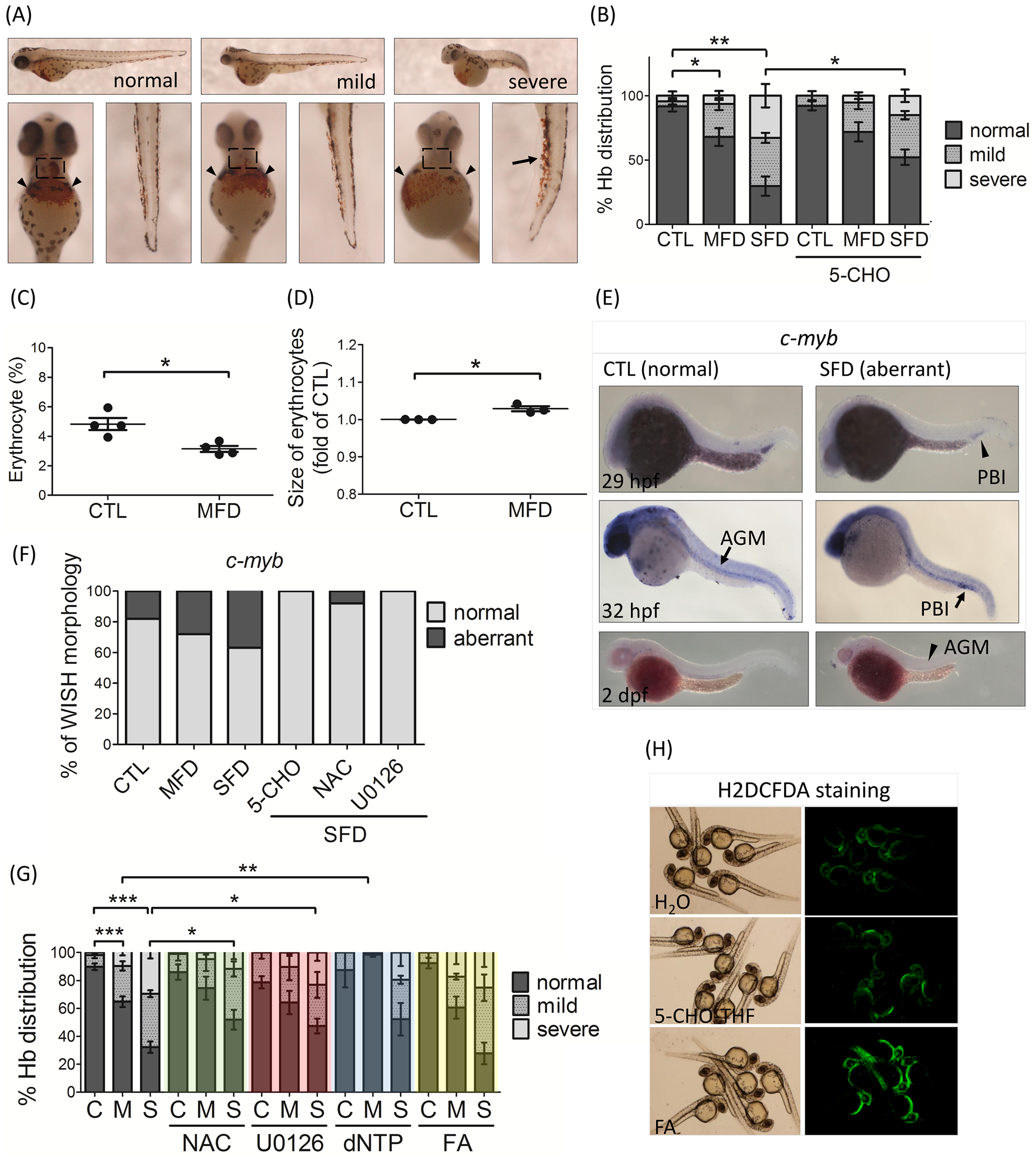
Fig. 3
Zebrafish larval hematopoiesis and response to rescuing agents.
(A, B) Hemoglobin of larvae in control and FD groups, with/without folate supplementation, were stained with o-dianisidine at 3 dpf. Hemoglobin signals were distributed most abundantly in the heart (dashed rectangles) and common cardinal veins (arrowheads) of control larvae (normal). Ectopic accumulation of hemoglobin in caudal veins (arrows) was often observed in FD larvae (mild and severe). The severity of anomalies was categorized and quantified based on the level and distribution of hemoglobin signals in larval heart and common cardinal veins. The images shown were the lateral (the upper panel) and ventral (the lower panel) views of larvae. Average of at least six independent experiments with the total sample number of 51–139 for each group are reported. (C, D) The relative number and size of embryonic erythrocytes were analyzed with flow cytometry for both control and FD embryos of 2-dpf generated by crossing Tg (hsp:EGFP-γGH) and Tg (gata1:dsRed). The numbers of erythrocytes were presented as the percentage of red fluorescent cells to total cell number. The size of erythrocytes was normalized with those of control larvae. Presented are data collected from at least three independent experiments with a total embryo number of approximately 30–40 for each group. (E) Hematopoiesis in both control and FD embryos was characterized by whole mount in situ hybridization with a riboprobe specific to c-myb, a hematopoietic stem cells marker. Reduced signals (arrowheads) with spatially and temporally altered distribution (arrows) were observed in embryos with severe folate deficiency. The larval responses to rescuing agents were quantified based on the distribution patterns of the c-myb signal at 32 hpf larvae (F) as shown in (E), and on the hemoglobin level (G) as shown in (A). There were approximately 10 to 40 larvae included for each group. (H) The 1-dpf wild-type larvae exposed to folic acid or 5-CHO-THF for 1 hour were examined for oxidative stress with H2DCFDA staining. C or CTL, heat-shocked non-fluorescent transgenic control; M or MFD, mild folate deficiency; S or SFD, severe folate deficiency; 5-CHO, 5-formyltetrahydrofolate; NAC, N-acetyl-L-cysteine; FA, folic acid. *, p<0.05; **, p<0.01; ***, p<0.001.