
|
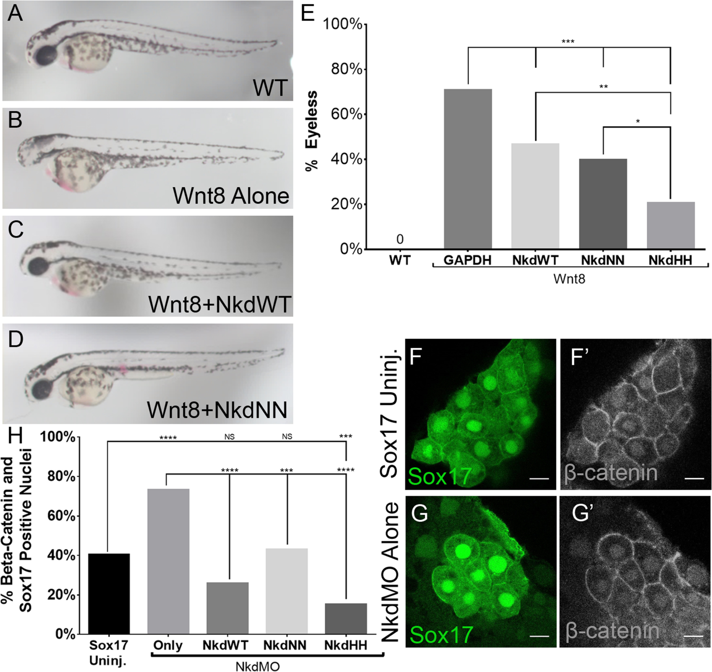
Fig. 3
NkdWT, NkdNN, and NkdHHantagonize Wnt/β-catenin. (A-D) Live images of 28hpf embryos, lateral view, anterior to the left. (A) Wild-type, (B) Wnt8+GAPDH RNAs, (C) Wnt8+NkdWT RNAs, and (D) Wnt8+NkdNN RNAs. (E) Graph of percentage of embryos without eyes. hpf, hours post fertilization. Uninj n=230, Wnt8 alone n=148, Wnt8+GAPDH n=34, Wnt8+NkdWT n=71, Wnt8+NkdNN n=44, Wnt8+NkdHH n=53. ***=p-value<0.0001, **=p-value<0.001, Fishers Exact Test. (F-G’) Confocal images of DFCs stained with anti- β-catenin. Nkd Antagonizes β-catenin Nuclear Localization in the DFCs. (F and G) Sox17-GFP which marks the DFCs. (F’ and G’) anti-β-catenin staining. (F-F’) Uninjected Sox17. (G-G’) NkdMO injected transgenic Sox17 line. (H) Graph showing percent (%) of embryos with β-catenin and Sox17 positive nuclei. Note that all forms of Nkd were able to rescue morphantsto uninjected levels, and that NkdHH was the most robust. Sox17 uninj. n=211, NkdMO alone n=134, NkdMO+GAPDH n=153, NkdMO+NkdWT n=70, NkdMO+NkdNN n=77, NkdMO+NkdHH n=99. n=number of cells. ****=p-value<1×10⁻⁵, ***=p-value<0.0001, Fishers Exact Test. Scale bar 10 µm. Confocal images taken at 63x at 2x zoom. See also Fig. S5.
Reprinted from Developmental Biology, 434(1), Marsden, A.N., Derry, S.W., Schneider, I., Scott, C.A., Westfall, T.A., Brastrom, L.K., Shea, M., Dawson, D.V., Slusarski, D.C., The Nkd EF-Hand Domain Modulates Divergent Wnt Signaling Outputs in Zebrafish, 63-73, Copyright (2017) with permission from Elsevier. Full text @ Dev. Biol.