Fig. 6
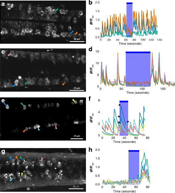
Calcium imaging of spinal neurons in GtACR1 embryos and non-expressing siblings. a Ventral spinal neurons of a 24-h-old 1020:GAL4, UAS:GtACR1-eYFP, elavl3:GCaMP6f embryo. The green arrowhead indicates a neuron with bright membrane label and puncta, suggesting expression of GtACR1-eYFP. The orange arrowhead indicates a neuron without GtACR1-eYFP expression. b Time course of fluorescence intensity in neurons, represented as change relative to minimum fluorescence. There is a reduction in activity during delivery of blue light. c, d Activity in dorsal spinal neurons, which do not express the GAL4 driver, in another 24-h-post-fertilization (hpf) GAL4s1020t, UAS:GtACR1-eYFP, elavl3:GCaMP6f embryo. Spontaneous activity was detected before and after, but not during, the period of blue light delivery. e, f Spontaneous activity in six neurons in a 26-h-old embryo with no GtACR1-eYFP expression. e Increase in fluorescence intensity occurs during the period of illumination with blue light (arrowheads). g, h The response of six spinal neurons in a 4-day-old embryo expressing GtACR1. There is no activity before light in these neurons, but there is a rise in GCaMP6f fluorescence after termination of the blue light. In panels b, d, f and h, the blue shaded region and bar indicate the period in which light was delivered. The colours of the traces represent relative change in fluorescence of the cells indicated by the arrowheads with the corresponding colours in the image on the left

