Fig. 5
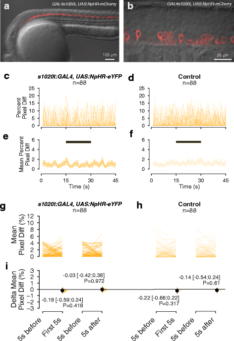
The effect of low intensity amber light on spontaneous movement of NpHR-expressing embryos. a, b Lateral view of a 24-h-old embryo expressing NpHR-mCherry under the 1020 GAL4 driver. a Overview of expression in the spinal cord. b. High magnification view, showing expression of NpHR-mCherry in spinal neurons. c–f Time course of movement of embryos, with (c, e) or without (d, f) halorhodopsin expression, in a 45-s recording with exposure to 15 s of amber light. The period of illumination is indicated by the black bar. c and d show movement of individual embryos, while e and f show mean values and 95% CIs. g, h Average amount of movement in the 5 s before light onset (‘5 s before’), in the first 5 s after light onset (‘First 5 s’) and in the first 5 s after light offset (‘5 s after’), in NpHR-expressing embryos (g) and non-expressing siblings (h). i Difference in movement in the first 5 s after light onset and after light offset, relative to the period before light, in NpHR-expressing and non-expressing embryos. Intensity of amber light used = 17 μW/mm2

