
|
Fig. 1
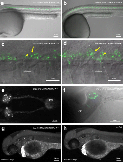
Expression of GtACR1 and GtACR2 in transgenic zebrafish. a, b Expression of GtACR1-eYFP in the trunk of 1-day-old GAL4s1020t, UAS:GtACR1-eYFP (a) and GAL4s1020t, UAS:GtACR2-eYFP (b) embryos. Labelled cells are located in the ventral regions of the spinal cord. c, d High magnification view of the trunk of fish expressing GtACR1-eYFP (c) or GtACR2-eYFP (d) in spinal neurons. There is label in the plasma membrane (arrows). For GtACR2, the label is dimmer. Puncta are visible (arrowheads). e An 8-dpf gng8:GAL4, UAS:GtACR1-eYFP larva, showing label in habenula neurons and in axons innervating the interpeduncular nucleus. f Expression of GtACR1-eYFP in olfactory neurons of a 3-day-old fish carrying the GAL4s1011t driver. g, h Forty-eight-hour-old embryos labelled with acridine orange. Dying cells are strongly labelled and appear as bright spots. These are detected in both GtACR1-expressing (g) and non-expressing (h) fish. There is no evidence of cell death in the ventral spinal cord of GtACR1-expressing fish. Anterior is to the left in all panels except f, where anterior is to the top. All panels are lateral views, except e and f, which are dorsal views. Panels a, c, d and f are single planes, while b, e, g and h are maximum projections. OE olfactory epithelium, IPN interpeduncular nucleus, lHb left habenula, rHb right habenula