
Image
|
Figure Caption
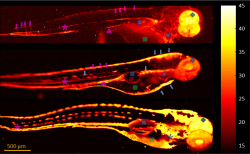
Fig. 4 AM comparison between 120 hpf larvae from differently pigmented zebrafish strains using fast scanning PAM. The color bar shows the SNR in decibels. Upper image, mitfa692/692, ednrba140/140 (AB) zebrafish, PTU treated; middle image, wild type AB* zebrafish, PTU treated; lower image, Tg(Fli:GFP) zebrafish. Blue arrows, pigment; blue square, swim bladder; blue diamond, eye; blue star, heart; magenta star, dorsal aorta; tip of magenta triangle, axial vein; magenta arrows, intersegmental vessels; green square, yolk sac.
Acknowledgments
This image is the copyrighted work of the attributed author or publisher, and
ZFIN has permission only to display this image to its users.
Additional permissions should be obtained from the applicable author or publisher of the image.
Full text @ Opt. Lett.