
Image
|
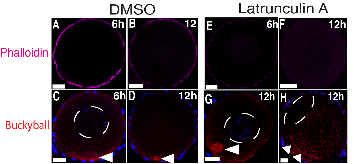
Figure Caption
Fig. S1
Latrunculin A treatment of stage I oocytes.
Ovaries treated with DMSO (A-D) or LatA (E-H) for 6h or 12h, then fixed and stained with phalloidin (magenta) (A, B, E and F) or Buc (red) (C, D, G and H). Arrowheads point to Buc localized to the Bb and the cortex. After 6h of treatment, no effect was found on the Bb or nucleus in 18 DMSO or 22 LatA treated oocytes. After 12h of treatment, 19 DMSO-treated oocytes were normal, whereas 4/22 LatA-treated oocytes showed Buc cortical detachment, three of which showed an acentric nucleus. N ≥ 5 ovaries. Scale bar: 20 μm.
Acknowledgments
This image is the copyrighted work of the attributed author or publisher, and
ZFIN has permission only to display this image to its users.
Additional permissions should be obtained from the applicable author or publisher of the image.
Full text @ PLoS Genet.