
|
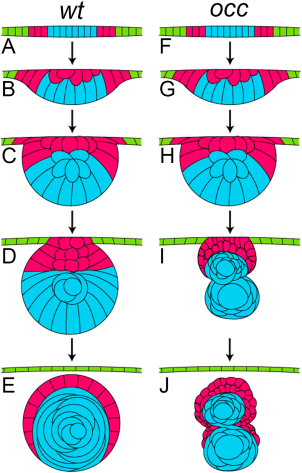
Fig. 8
Comparison of the developmental stages for lens in wt and occ Zebrafish. In this diagram, the developmental stages of the lens in a wt eye (A–E) were compared with the developmental stages of the lens in the eye of an occ mutant (F–J). In the early developmental stages, the lens cell mass delaminated normally from the surface ectoderm in the wt and occ eyes, and the lens epithelial cells began differentiating into secondary lens fibers (A–C, F–H). In the occ eye, the multilayered epithelial cells formed a secondary mass of cells anterior to the surface of the developing lens (I). By 4 dpf, the wt lens formed a core of well-defined nuclear cells surrounded by an epithelium and continued to develop normally to become a functioning adult lens (D,E). In the occ eye, a small secondary cellular mass became ordered and multilayered as the other lens degenerated (I,J). With further development, the secondary mass of epithelial cells in the occ mutant differentiated into the lens fiber cells (J). The abnormal development of the occ lens accounted for the abnormal visual function in the occ mutant. While there was variability, the drawings represent the developmental stages observed using live-embryo imaging and histology. (See Suppl. Movies 1–3.)