
Image
|
Figure Caption
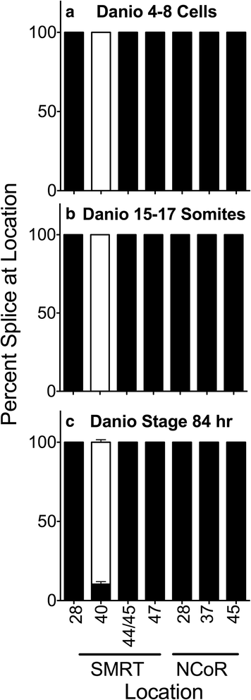
Fig. 3 Alternative splicing during Danio rerio development. Entire individuals were isolated at the stages indicated, RNA was extracted, and the alternative splicing at the indicated locations was analyzed and is presented graphically as in Fig. 2. Mean and standard deviation are shown (n = 3) for each. Panels represent Danio 4-8 cell stage animals (a), Danio 15-17 somite stage animals (b) and Danio stage 84 hour animals (c)
Figure Data
Acknowledgments
This image is the copyrighted work of the attributed author or publisher, and
ZFIN has permission only to display this image to its users.
Additional permissions should be obtained from the applicable author or publisher of the image.
Full text @ BMC Evol. Biol.