
|
Fig. S8
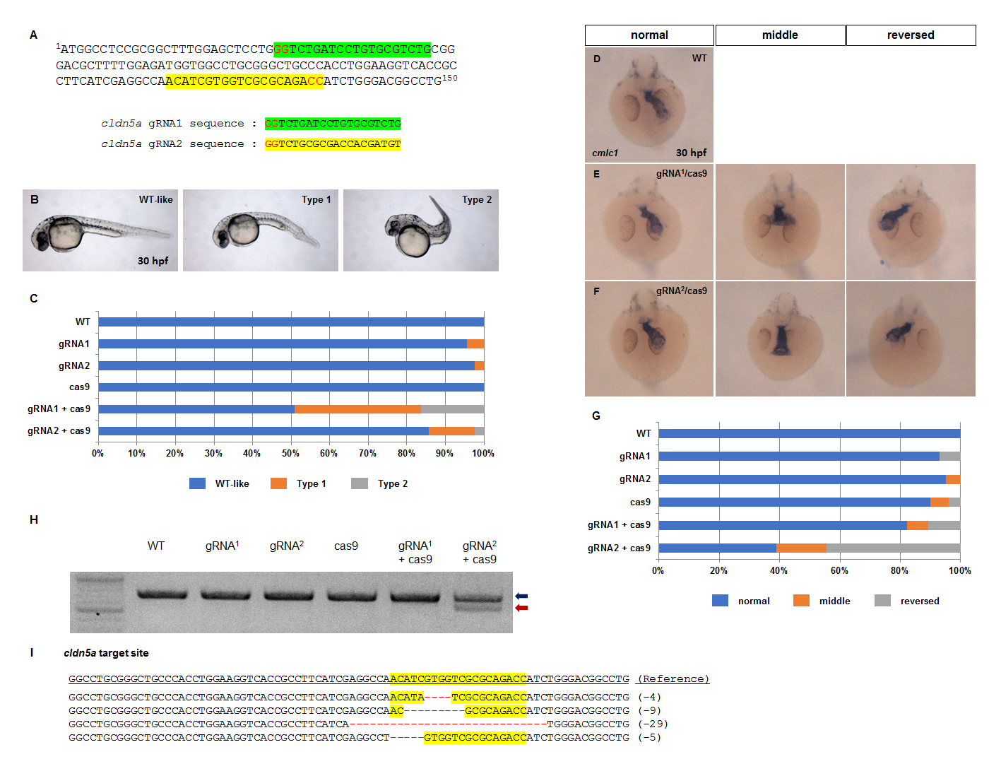
Laterality of heart was disrupted in cldn5a crispants.
(A) Partial nucleotide sequences of cldn5a coding sequence (1–150 among 648) and two types of cldn5a targeting gRNA sequences. (B) Representative images of WT-like, type1 and type2 embryos at 30 hpf. (C) Stacked bar graph (blue; WT-like, orange; type1, grey; type2, WT; n = 24, 40 pg of gRNA1 injected embryos; n = 45, 40 pg of gRNA2 injected embryos; n = 41, 80 pg of cas9 mRNA injected embryos; n = 52, 40 pg of gRNA1 and 80 pg of cas9 mRNA injected embryos; n = 55, 40 pg of gRNA2 and 80 pg of cas9 mRNA injected embryos; n = 42). (D—F) Visualization of a heart by in situ hybridization of cmlc1 in 30 hpf embryos. Representative images of WT (F), gRNA1 crispants (G), and gRNA2 crispants (H). (G) Stacked bar graph (blue; normal, orange; middle, grey; reversed, WT; n = 24, only gRNA1 injected embryos; n = 43, only gRNA2 injected embryos; n = 40, only cas9 mRNA injected embryos; n = 52, gRNA1 crispants; n = 28, gRNA2 crispants; n = 36). (H) T7E1 analysis of cldn5a crispants. (I) Representative mutations of cldn5a gene in gRNA2 crispants.