
Image
|
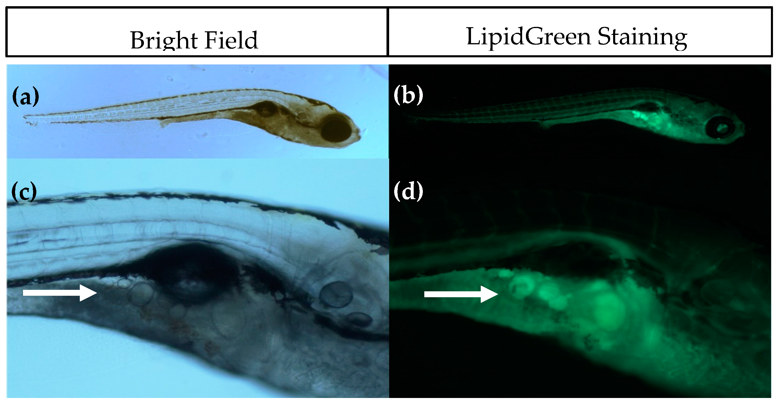
Figure Caption
Fig. 1
Imaging of adipocytes using Bright Field (left) and fluorescence microscopy after LipidGreen staining (right). (a,b) Representative images of a 15 days post fertilisation (dpf) larvae (5× magnification); (c,d) Close-up of the pancreatic area where the first adipocytes develop. Adipocytes are indicated with a white arrow (16× magnification).
Acknowledgments
This image is the copyrighted work of the attributed author or publisher, and
ZFIN has permission only to display this image to its users.
Additional permissions should be obtained from the applicable author or publisher of the image.
Full text @ Int. J. Mol. Sci.