
|
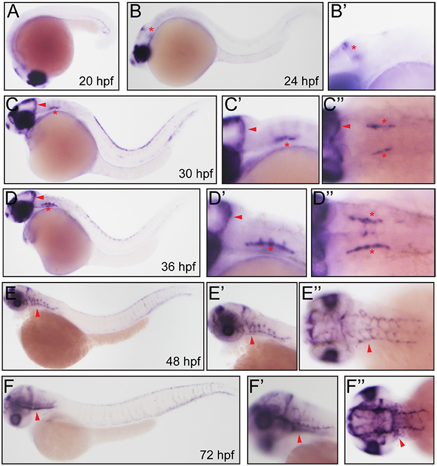
Fig. 2
Whole mount in situ hybridization analysis of fabp11a in zebrafish embryos. (A) 20 hpf, lateral view. (B) 24 hpf, lateral view. Red asterisk indicates mid-cerebral vein (MCeV). (B') 24 hpf, lateral view. Red asterisk indicates mid-cerebral vein (MCeV). (C) 30 hpf, lateral view. Red arrowhead indicates MCeV; Red asterisk indicates primordial hindbrain channel (PHBC). (C') 30 hpf, lateral view. Red arrowhead indicates MCeV; Red asterisk indicates PHBC. (C'') 30 hpf, dorsal view. Red arrowhead indicates MCeV; Red asterisks indicate PHBC. (D) 36 hpf, lateral view. Red arrowhead indicates MCeV; Red asterisk indicates primordial hindbrain channel (PHBC). (D') 36 hpf, lateral view. Red arrowhead indicates MCeV; Red asterisk indicates PHBC. (D'') 36 hpf, dorsal view. Red arrowhead indicates MCeV; Red asterisks indicate PHBC. (E,E') 48 hpf, lateral view. Red arrowhead indicates brain vessels. (E”) 48 hpf, dorsal view. Red arrowhead indicates brain vessels. (F,F') 72 hpf, lateral view. Red arrowhead indicates brain vessels. (F'') 72 hpf, dorsal view. Red arrowhead indicates brain vessels.