
Image
|
Figure Caption
Fig. S3
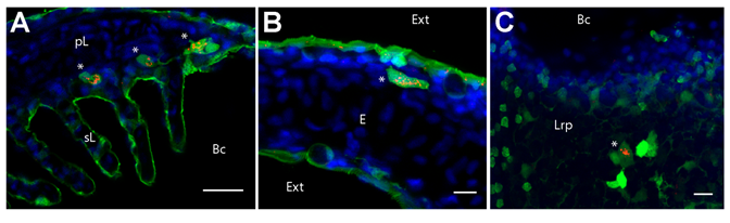
Cells with high intracellular and granular PNA staining express MHC2. Confocal images of gill (A), skin (B) and thymus (C) cryosections from mhc2dab:GFP adult fish, following PNA (red) and DAPI (blue) staining. Single optical section (A) or maximal intensity projections from 9 (B) or 5 (C) optical sections taken every micrometer. pL (primary Lamellae), sL (secondary Lamellae), Bc (Branchial cavity), Ext (External environment), E (Epidermis), Lrp (Lympho-reticular parenchyma). Scale bar: 10 µm (B, C), 20 µm (A).
Acknowledgments
This image is the copyrighted work of the attributed author or publisher, and
ZFIN has permission only to display this image to its users.
Additional permissions should be obtained from the applicable author or publisher of the image.
Full text @ Front Immunol