
|
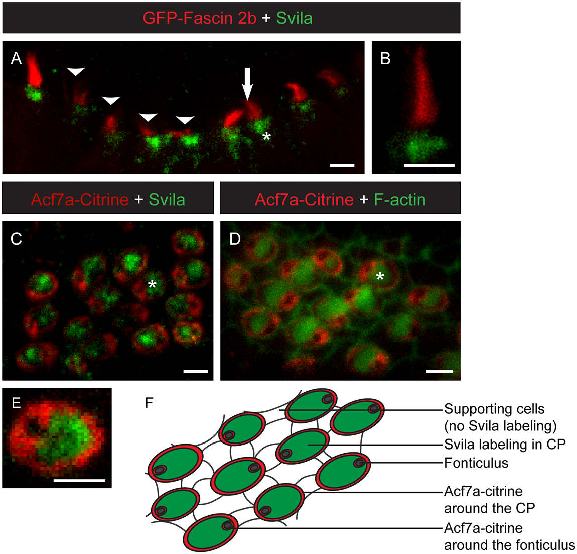
Fig. 4
Supervillin localizes to zebrafish hair cell CPs.
(A,B) Confocal micrographs of 4-dpf zebrafish anterior crista hair cells expressing GFP-fascin 2b (red) and labeled with anti-Svila (green). Arrowheads in A indicate hair cells out of focus or bent. Arrow indicates hair-bundle-localized GFP-fascin 2b. Asterisk indicates a CP. (C,D,E) Confocal micrographs of posterior macula hair cells from zebrafish expressing Acf7a-Citrine (red) and labeled with anti-Svila (green) (C,E) or phalloidin (green) (D). Asterisks in (C,D) indicate CPs. Acf7a-Citrine encircles the CP, localizes to the CP base, out of the focal plane in C-E, and is found weakly throughout the CP. (F) Schematic of the zebrafish posterior macula tissue with the location of Svila immunolabeling in green and Acf7a-Citrine indicated in red. Scale bars, 2 μm.