
|
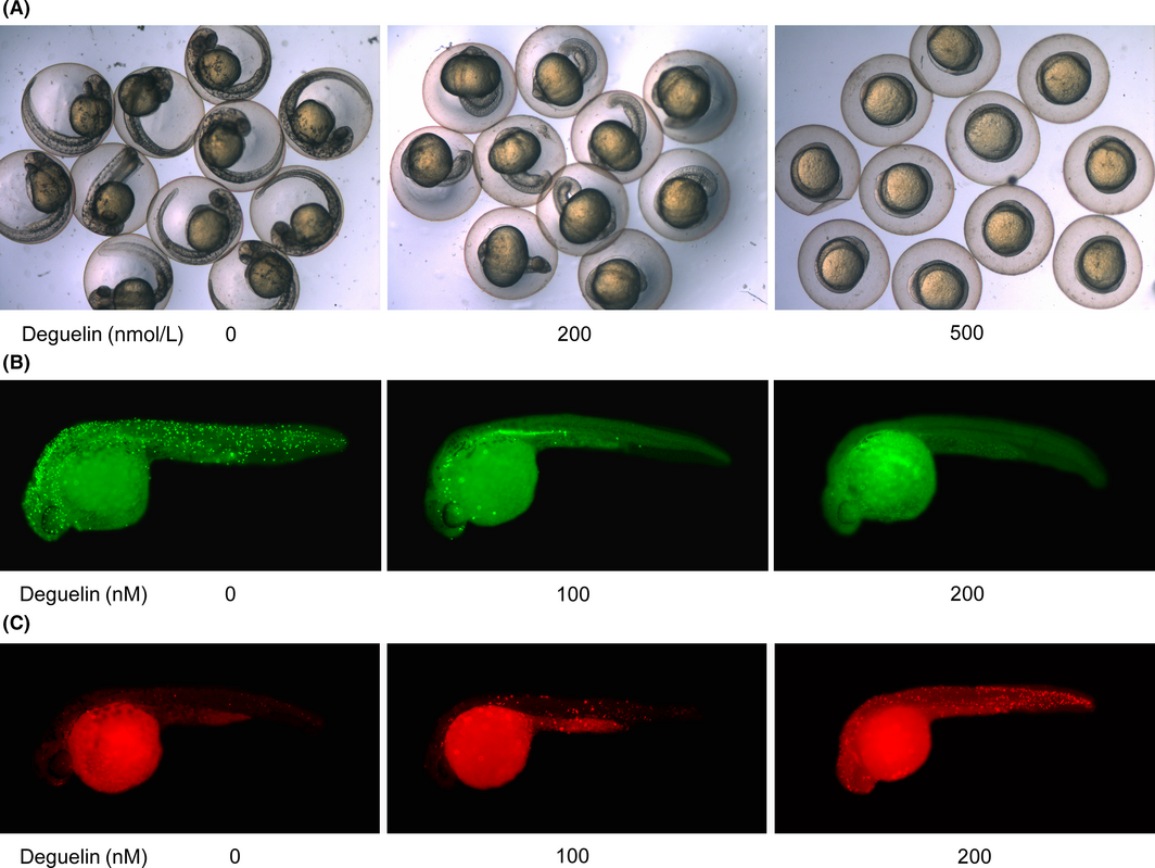
Fig. 1
Growth repression and apoptosis induction caused by deguelin. (A) Morphological change in zebrafish with or without deguelin treatment. Significant growth retardation can be found in 200 and 500 nmol/L deguelin-treated group. (B) Whole-mount embryos labeled with anti-pH3 antibody to examine proliferating cells in zebrafish larvae. The numbers of pH3-positive cells decreased dramatically and rarely expressed with 200 nmol/L deguelin treatment (magnification 50×). (C) Phenotypic assessed by terminal deoxynucleotidyl transferase dUTP nick end labeling (TUNEL) staining. There was a dose-dependent increase of apoptotic cells in TUNEL assay. (magnification 50×).