
Image
|
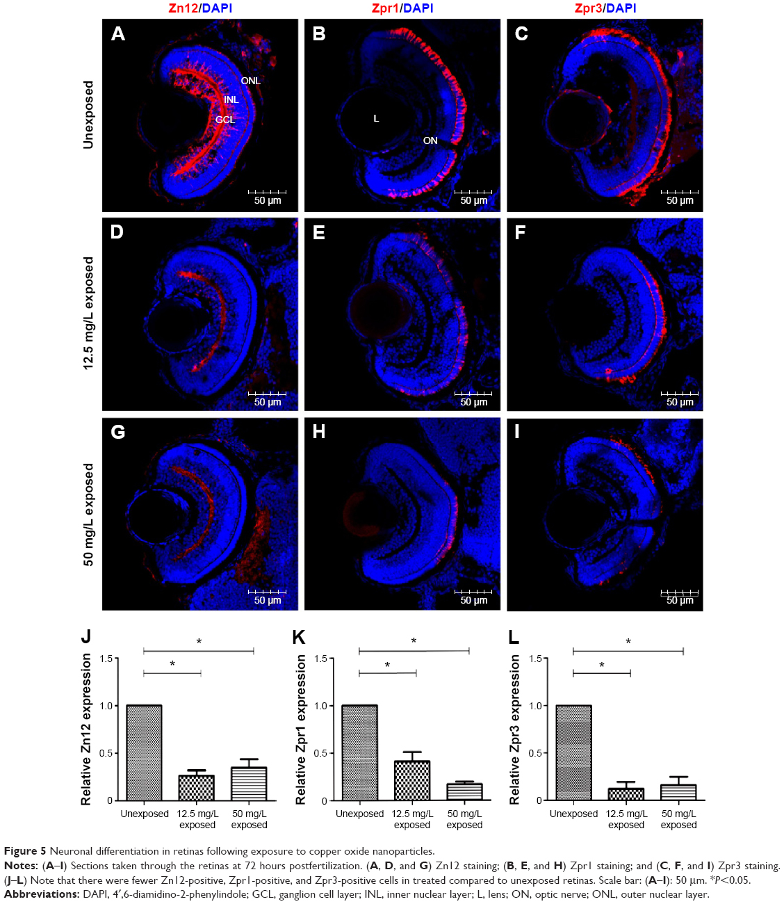
Figure Caption
Fig. 5
Neuronal differentiation in retinas following exposure to copper oxide nanoparticles.
Notes: (A–I) Sections taken through the retinas at 72 hours postfertilization. (A, D, and G) Zn12 staining; (B, E, and H) Zpr1 staining; and (C, F, and I) Zpr3 staining. (J–L) Note that there were fewer Zn12-positive, Zpr1-positive, and Zpr3-positive cells in treated compared to unexposed retinas. Scale bar: (A–I): 50 μm. *P<0.05.
Abbreviations: DAPI, 4′,6-diamidino-2-phenylindole; GCL, ganglion cell layer; INL, inner nuclear layer; L, lens; ON, optic nerve; ONL, outer nuclear layer.
Acknowledgments
This image is the copyrighted work of the attributed author or publisher, and
ZFIN has permission only to display this image to its users.
Additional permissions should be obtained from the applicable author or publisher of the image.
Full text @ Int. J. Nanomedicine